WHAT IS THE CARTILAGE IN THE KNEE FOR?
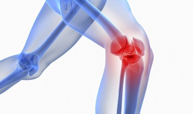
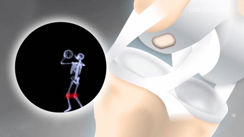
An approx. 5 mm thick layer of cartilage covers the bone in the knee joint. The smooth surface of the cartilage allows the joint to move smoothly. In addition, the cartilage causes an even distribution of pressure and shock loads on the underlying bones. This protects the bone and prevents or reduces overstressing. The cartilage has no nerves and stops growing after puberty. This layer of cartilage accompanies us throughout life.
HOW DOES CARTILAGE DAMAGE COME?
The cartilage undergoes natural aging. In doing so, it loses the ability to store water and the cartilage layer shrinks. The surface of the cartilage becomes brittle and cracks. This brittle surface is more prone to impacts and shear forces. This allows the cartilage to wear out or split off more quickly. Since the cartilage has no pain fibers, we do not feel this change at the beginning. Perhaps a rubbing noise (crepitation) is noticeable under greater stress. In contrast to cartilage, bone has pain fibers. If the bone is exposed, we feel the affected joint with the corresponding pain, explains the orthopaedic in Delhi.
However, the articular cartilage can also be damaged by chronic stress or an accident (trauma).
HOW DO YOU RECOGNIZE CARTILAGE DAMAGE?
With acute cartilage damage, patients complain of blockage of the knee joint, swelling and pain. The extent of the discomfort depends on the size and depth of the cartilage defect and its location.
In the case of chronic cartilage damage, patients report start-up, stress, and inflammatory pain. The knee is swollen, and the mobility of the knee joint is limited. In addition, the patients have an unsteady gait, the knee joint feels unstable and in some cases kinks, says the orthopaedic in Delhi.
HOW IS CARTILAGE DAMAGE DIAGNOSED?
The damage to the articular cartilage can appear superficially with small cracks on the one hand, but on the other hand affect the entire cartilage in the knee. This causes the rough, painful surface of the bone to emerge.
Cartilage damage is divided into four stages:
- Stage / Grade I: Soft cartilage
- Stage / Grade II: Rough surface with cracks
- Stage / Grade III: Deep cracks in the cartilage that go down to the bone
- Stage / Grade IV: Complete consumption of the cartilage with exposed boil; one also speaks of bald bones
In addition to the exact questioning (anamnesis) and well-founded clinical examinations of the knee joint, X-ray and MRI images are necessary. Based on these documents, the appropriate individual therapy can be discussed, says the orthopaedic doctor in Delhi.
WHAT TREATMENTS ARE AVAILABLE FOR CARTILAGE DAMAGE?
The cartilage damage looks different depending on the cause, whether accidental (acute) or wear-related (chronic) and is treated differently accordingly. In the case of acute cartilage damage, for example, we have a clearly defined defect (punch defect) compared to healthy cartilage with sharp edges. This is not the case with chronic cartilage defects. If the cartilage defect is not treated, there is further cartilage wear of the knee joint and, in the further course, knee joint osteoarthritis.
Conservative therapy for cartilage damage is very limited. After puberty, the cartilage loses its self-healing potential, i.e. from this point onwards we have to get by with the cartilage for our entire life. For these reasons, the natural course of cartilage damage leads to deterioration. The cartilage damage gets bigger and deeper over time, which leads to a clinical deterioration with corresponding pain and restrictions in everyday life and during sporting activity. With conservative therapy, cartilage damage cannot be cured, but only alleviated by slowing down the wear and tear of the cartilage. The following conservative therapies are possible:
- physical therapy
- Osteopathy
- Painkiller
- Cartilage Support Agents
- Lubricating syringes: Hyaluronic acid syringes temporarily improve the joint lubrication of the defective joint. This will reduce the inflammation in the joint and, accordingly, the patient will experience less pain. Injections with autologous blood are another option.
- More: Pay attention to your own weight or reduce it, a healthy balanced diet, regular training with guided movements such as swimming or cycling.
The surgical therapy of the cartilage damage depends on the size (extent) and depth of the defect and must be individually adapted. In addition, factors such as the integrity of the exposed bone, cartilage quality on the opposite side of the defect and the age of the cartilage defect play a decisive role, explains the orthopaedic surgeon in Delhi.
Furthermore, the younger the patient, the greater the chances of success of the methods described below for acute cartilage damage. These include:
- Fixation of the bone / cartilage splinter with a mostly dissolvable screw.
- Arthroscopic sealing of superficial cartilage tears.
- Microfracture: First, the cartilage defect is prepared in such a way that a clean, stable cartilage margin is present. Then small holes are made in the exposed bone with the drill or special awls. It is from these holes that blood comes out of the bone, which contains stem cells. Among other things, these stem cells can transform into cartilage and thus protect the exposed bones again. This method is mostly used arthroscopically (joint endoscopy) and is suitable for smaller cartilage defects (<2.5 cm2).
- Transplantation of cartilage-bone cylinders: This technique is also called mosaic plastic or OATS (Osteochondral Autologous Transplantation System). The method is also only used for smaller cartilage defects (<2.5 cm2). With special hollow punches, the body's own cartilage-bone cylinders are taken from an unloaded part of the knee joint and inserted directly into the cartilage defect. This method is used when, in addition to the cartilage defect, the underlying bone is also damaged.
- AMIC: The AMIC technique is used for larger cartilage defects. AMIC stands for autogenous matrix-induced chondrogens, i.e. matrix-induced own cartilage production. The cartilage defect is exposed and cleaned so that stable cartilage edges are present again. This is followed by micro-invoicing as described above.
- Cartilage transplantation: Another option would be to have your own cartilage cells grown in a laboratory from biopsies obtained from the first operation. This method is also called ACT (autologous chondrocyte transplant). After about four weeks of cultivation in a laboratory, the cartilage cells can be inserted into the corresponding defect in a second operation.
- Bone defects: If there are bone defects that are deeper than 4 mm, they must be filled with the body's own bone (e.g. thigh) or donor bones (cancellous plastic).
The above-mentioned surgical techniques are mainly used for acute cartilage defects. In addition to age, the opposite side of the cartilage defect also plays a role. This should not show any major damage, otherwise the rough surface can negatively affect the outcome of the operation. Another option for covering more chronic cartilage defects is knee replacement in Delhi of the injured cartilage.
If the cartilage wear has progressed so far that finally bone rubs on bone (end stage of osteoarthritis), a partial prosthesis (e.g. a sled prosthesis, a kneecap glider replacement) or a knee replacement surgery in Delhi is necessary. A correction of bowlegs / knock knees (so-called corrective osteotomy) is sometimes necessary.
WHAT IS THE AFTER-TREATMENT LIKE?
The follow-up treatment must be individually adapted to the previous operation. If cartilage therapy was carried out, the knee joint had to be relieved with walking sticks for six weeks. In the case of cartilage defects behind the kneecap, mobility is also restricted for six weeks. Patients with resurfacing can immediately put weight on and move the knee joint. To protect the soft tissues, however, walking sticks are recommended for around four weeks. Physiotherapy for several weeks to relieve swelling and strengthening and stretching of the thigh muscles are also important.