Showing posts with label orthopaedic doctor in Dwarka. Show all posts

Saturday, 14 September 2024

When to Visit an Orthopaedic Specialist


Orthopaedic conditions can significantly affect your daily life. Recognizing when to see a specialist can help prevent further damage and improve your quality of life. Here are some key signs to help you decide when it’s time to consult an orthopaedic surgeon in Dwarka.

Chronic Pain in Joints or Bones

If you experience persistent pain in your joints or bones lasting for more than a few weeks, it may be a sign of an underlying condition. Acute injuries often heal with rest, but if discomfort continues, an orthopaedic in Dwarka can diagnose the cause and recommend treatment.

Limited Range of Motion

A decreased ability to move your joints fully is another clear indication to visit an orthopaedic in Dwarka. Limited range of motion can result from injuries, arthritis, or other degenerative conditions. Early intervention can help restore mobility and prevent the issue from worsening.

Injuries from Sports or Accidents

Sports-related injuries, car accidents, or slips and falls often lead to sprains, fractures, or dislocations. If you’ve sustained an injury that hasn’t healed properly or you’re unsure of the severity, an orthopaedic consultation is essential for a thorough evaluation and effective recovery plan.

Difficulty in Performing Daily Activities

If everyday tasks like walking, climbing stairs, or lifting objects become challenging due to joint or bone pain, it’s time to seek professional help. An orthopaedic doctor in Dwarka can identify the cause and provide solutions such as physical therapy, medications, or even surgery if necessary.

Swelling and Inflammation

Unexplained swelling, redness, or inflammation in the joints or around the bones is a warning sign that something may be wrong. These symptoms could indicate an infection, injury, or inflammatory disease like arthritis. An orthopaedic doctor in Dwarka can help pinpoint the cause and recommend appropriate treatments.

Bone Deformities or Abnormal Growths

Visible deformities or abnormal growths in the bones, whether from injury or a congenital condition, should be evaluated by an orthopaedic doctor in Delhi. These issues can impact your physical appearance and functionality, making early diagnosis critical for managing symptoms effectively.

Back Pain That Persists

Chronic back pain is one of the most common reasons people visit orthopaedic doctor in Delhi. If over-the-counter pain relievers and rest haven’t alleviated the issue, it’s a good idea to consult a specialist. They can assess whether the pain is due to a muscle strain, spinal issue, or something more serious.

Numbness, Tingling, or Weakness in Limbs

Experiencing numbness, tingling, or weakness in your arms or legs could be related to nerve compression or a spine issue. These symptoms should not be ignored as they can worsen over time, leading to permanent damage if left untreated.

Previous Surgeries with Lingering Symptoms

If you’ve had surgery on your bones or joints but still experience lingering symptoms, it’s worth a visit to the orthopaedic surgeon in Dwarka for a follow-up. They can assess whether the initial procedure was successful or if further treatment is necessary to fully address the problem.

Conclusion

Orthopaedic issues can affect anyone at any age. If you experience any of the symptoms mentioned, don’t hesitate to consult a specialist. Early detection and treatment can make a significant difference in your recovery and long-term health.

Wednesday, 17 August 2022

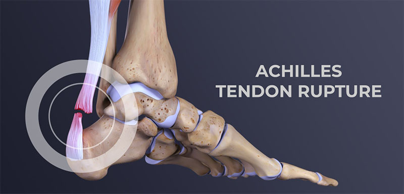
Achilles tendon rupture - treatment, surgery


What is Achilles tendon rupture?

An Achilles tendon rupture or rupture occurs when the tendon “ruptures or tears” leading to separation or discontinuity in the tissues that make up the tendon.

In terms of anatomy, the Achilles tendon, a sort of fibrous “ribbon” or “cord” that connects muscle to bone, is the largest and strongest tendon in the body that connects the calcaneus (heel bone) to the medial gastrocnemius muscles., lateral gastrocnemius and soleus muscles (popularly known as calf muscles or “leg belly” muscles). When the muscle contracts, it “pulls” the tendon, which in turn moves the foot.

The rupture often reflects the previous existence of tendinosis phenomena caused by sports microtraumas or degeneration (aging) of the tendon. In most cases, the tear occurs at the bottom of the tendon (just above the heel), but it can occur anywhere along the tendon. In the presence of a healthy tendon, the lesion can appear in the bone or muscle.

Total ruptures, partial ruptures

We can generically classify Achilles tendon ruptures into:

  • Total rupture – in the total rupture of the Achilles tendon, the tissues break completely, that is, the tendon is completely “separated”.
  • Partial rupture – in a partial rupture of the Achilles tendon, the tendon does not completely rupture, only an incomplete rupture occurs. Partial tears can vary greatly in severity according to the extent of the injury.

A total rupture is more frequent than a partial rupture. As a rule, a total rupture is a more serious injury when compared to a partial rupture and with more exacerbated symptoms. A partial tear can also present with pain that can range from moderate to severe and, if not recognized, it can quickly progress to a total tear.

See more information on treatments to better understand the different therapeutic approaches according to the extent of the lesion.

Achilles tendon rupture – causes

The Achilles tendon can lose elasticity and become “weak and thin” with age and lack of use. Then, it becomes prone to injury or breakage.

Rupture is more common in people with pre-existing Achilles tendonitis (tendon inflammation). Repetitive tendinitis and the consequent calcifications are a risk factor for tendon rupture.

Certain diseases (such as arthritis and diabetes) and medications (such as corticosteroids, for example) can also increase the risk of rupture.

Rupture occurs most often in the middle-aged male athlete. The injury usually occurs during recreational sports that require impacts with the ground, running, jumping, etc. Most of the time, these are football, athletics, tennis, basketball, among others. Injury can happen in these situations as a result of traumatic dorsiflexion when the muscle is strongly contracted causing it to tear.

Breaks can, however, also occur in everyday activities. For example, when falling from a significant height, when entering a pothole abruptly, traffic accidents, etc.

Achilles tendon rupture – symptoms

The signs and symptoms of Achilles tendon rupture are:

  • Sudden, severe pain may be felt in the “back” of the ankle or the “tummy of the leg”, often described as “being hit by a rock or shot” or “like someone stepping on the back of the ankle”;
  • A loud clicking sound can be heard;
  • A discontinuity (“gap”) or depression (void) can be felt and seen in the tendon above the calcaneus (heel bone);
  • Inability to stand on tiptoe on the affected side;
  • Initial pain, swelling (swelling) and stiffness may be followed by bruising and weakness (not being able to land on the foot, walking).

Achilles tendon rupture – diagnosis

The diagnosis is made by the orthopaedic doctor in Delhi after collecting the clinical history, performing the physical examination and some complementary means of diagnosis (MCDT).Bottom of Form

A simple test is to “stretch” the “calf or calf muscles” while lying on your stomach (Thompson test). In the impossibility of being able to elevate the foot, there is, most likely, a rupture in the tendon. This test isolates the connection between the “calf muscles” and the tendon and eliminates other tendons that may still allow poor movement.

The orthopaedic doctor in Dwarka may order the following tests to confirm the diagnosis and to know in greater detail the location and degree of severity of the lesion:

  • Plain radiography (XR) – not being a very useful exam, it can identify a bone fragment avulsion of the calcaneus;
  • Ultrasound or ultrasound – Ultrasound of the leg and thigh can help assess the possibility of deep vein thrombosis and can also be used to rule out a Baker’s cyst (or cyst). Ultrasound can identify Achilles tendon rupture or signs of inflammation (tendinitis or tendinosis);
  • Magnetic Resonance Imaging (MRI) – MRI is extremely sensitive for diagnosis and allows you to determine if there is still a tendon in continuity. It allows other diagnoses such as tendinitis, tendinosis and bursitis.

Pain in the “back of the heel” is not always due to an Achilles tendon rupture. In the differential diagnosis, tendinitis (inflammation of the Achilles) and bursitis (inflammation of the bursae) should be considered, among the most frequent pathologies that cause pain in the Achilles region.

Achilles tendon rupture – treatment

The objective of the treatment is to restore the function of the tendon, for this, it is necessary that the tissues that make up the tendon heal “united” with each other. In this way, it will be possible for the patient to return to the same level of activity before the injury. Regaining Achilles tendon function after an injury is critical to making walking possible.

Treatment reflects a balance between tendon protection and initial movement. Protection is necessary to allow time for healing and to prevent further injury. Moving the foot and ankle is necessary to prevent stiffness and loss of muscle strength.

We can divide treatment options into surgical and non-surgical. Conservative (non-surgical) treatment consists of a set of therapeutic attitudes aimed at healing the tendon and restoring its function without resorting to any type of surgical intervention.

The choice between surgical and non-surgical treatment can be controversial in some cases. Both surgical and non-surgical treatment will require an initial period of approximately six weeks of immobilization. For most patients, both treatment options have good functional results.

NON-SURGICAL TREATMENT

Nonsurgical treatment is often used for non-athletes or for people with a low general level of physical activity who will not benefit from surgery. In the elderly and people with clinical complications, conservative (non-surgical) treatment should also be considered as a first option.

Initially, a cast below the knee is performed with the foot in equinus (foot in marked plantar flexion, “down”). Although it is not routine, it is possible to perform an MRI to verify that the tops of the tendon are in contact. The cast is changed, at intervals of two to four weeks, to slowly stretch the tendon back to its normal length. This treatment usually takes 8 to 12 weeks. During this period, global strengthening and flexibility exercises are taught.

SURGERY (SURGICAL TREATMENT)

Surgery on Achilles tendon rupture is often indicated in healthy and active people who want to resume activities such as walking, running, cycling, etc. Even those who are less active may be candidates for surgical repair of the tendon. The decision to operate should be discussed with your orthopaedic surgeon in Delhi.

Surgery should not be performed if there is an active infection or damaged skin at or around the Achilles tendon rupture site. In addition, some diseases or lifestyle habits, such as diabetes, smoking habits, sedentary lifestyle, steroid use and inability to follow instructions after the operation, may be a contraindication for surgery.

Surgical intervention for an Achilles tendon rupture is usually performed on an outpatient basis. This means that the patient is operated on and goes home the same day.

We can identify two distinct surgical approaches:

The first is to perform the intervention percutaneously, allowing to perform a minimally invasive surgery, through small incisions. A kind of needles with attached sutures are passed, allowing the Achilles tendon to be sutured.

The second approach is the open approach (traditional method, where the surgeon makes an open incision to access the tendon). This starts with an incision made in the back of the leg, just above the calcaneus (heel bone). After the best orthopaedic in Dwarka finds the two ends of the torn tendon, these ends are sutured. The incision is subsequently closed.

The surgical technique will be previously determined by the orthopaedic in West Delhi, depending on the type and location of the rupture, among other factors. In the postoperative period, regardless of the surgical technique chosen, the patient is immobilized with an equinus foot.

Despite being a safe surgery, some complications can arise, such as risks associated with anesthesia, infection, damage to nerves and blood vessels and bleeding or blood clots, among others. A new rupture can also occur (recurrence).

Recovery after surgery

After the surgery, the patient is placed with a splint or a “plaster boot” from the foot to the knee. Usually, the patient cannot walk or put weight on the involved leg. Crutches, a walker or a wheelchair are used to allow the patient to remain mobile for the first few times. Patients are encouraged to keep the operated leg elevated above the level of the heart to decrease swelling (swelling) and pain.

Patients are usually seen in the office two weeks after surgery. The splint or cast is removed, and the surgical incision is evaluated. Stitches are usually removed at this point if necessary. After two to six weeks, depending on postoperative protocol and orthopedic in Dwarka preference, patients may be allowed to begin performing some force. For this, a “hiking boot” can be used. Ankle movement is often allowed and encouraged.

After six weeks, full-body strength is generally allowed. Physiotherapy in Dwarka will need to be started and is intended to restore ankle range of motion. Strengthening of the “calf muscles” and Achilles is gradually allowed as the tendon heals. It is usually possible to resume full activity after six months. Recovery time after surgery can extend up to a year, until the patient can achieve full rehabilitation.

Even in cases where surgery is performed, the above-mentioned therapeutic attitudes are included in the rehabilitation plan. See more information on conservative treatment.

Monday, 18 July 2022

Hip Osteoarthritis Treatment

Hip arthrosis, also called coxarthrosis, belongs to a group of diseases called chronic degenerative diseases, that is, diseases that progressively evolve, affecting certain areas or tissues of the body.

In the case of osteoarthritis of the hip, there is progressive wear and tear of the cartilage of the hip joint.

This disease is also characterized by bone neoformation in the region where there was joint wear. These neoformed structures are popularly called parrot beaks.

Risk Factors for Hip Osteoarthritis

It is not known for sure why certain groups develop hip arthrosis, but it is known that some situations tend to increase the probability of developing this degenerative disease.

Among these factors are previous diseases of the hip, such as epiphysiolysis, in addition to septic arthritis, congenital dislocation, femoroacetabular impingement, rheumatism, fracture sequelae, as a result of previous surgeries, etc.

This pathology usually affects older people. In the case of previous diseases in the hip, it is called the pathology of secondary hip arthrosis. It is estimated that in the population over 65 years of age approximately 12% have symptomatic osteoarthrosis.

Symptoms

The main symptom of hip arthrosis is pain, located in the groin area. But the patient may report other symptoms such as difficulty performing simple movements such as bending down or bending, as well as joint stiffness and crepitus.

Other symptoms are pain in the buttocks area and the pain may be present even after a period of rest, especially at night.

Difficulty performing simple activities such as walking, climbing stairs, sitting or crossing the legs is present in cases of coxarthrosis.

As coxarthrosis is a progressive disease, the signs and symptoms also have a progressive evolution, that is, they can start in a very mild and little limiting way, but progress to intense and very limiting.

Thus, the first signs of coxarthrosis include joint stiffness, which starts bothering more in the morning, but tends to disappear during the day. In these cases, the limitation of movement is quite small.

With the passage of time and the evolution of the disease, joint involvement also increases and, as a result, the stiffness becomes greater and tends not to disappear during the course of the day. Even the pain can even radiate to other places, such as the lumbar spine, for example.

Rest no longer helps to improve the condition and the patient begins to feel pain in any location or position, feeling very uncomfortable even when lying down or standing still.

The consequence of this is that the patient increasingly decreases the level of movement and when he moves, he limps, trying to transfer the load to the other side of the body that does not present the pathology.

Although this seems like a temporary solution, it actually only worsens the situation, as it leads to muscle weakness in the leg and buttocks, which are extremely important musculature for hip protection.

Thus, it is important that a professional is sought when the first symptoms appear, so that the diagnosis is established and a good treatment plan made, precisely to prevent the natural progression of the disease from occurring.


Diagnosis of Osteoarthritis of the Hip

The diagnosis of this pathology is the responsibility of the orthopaedic doctor in Dwarka. Unfortunately, many patients are slow to seek medical help, believing that the pain and the situation will spontaneously improve, which is not the case with a degenerative disease.

Clinical evaluation is essential, with the professional collecting information about the pain history and medical history of that patient. In addition, some functional exams to check the patient's muscle capacity in the region are performed.

It is important to check the muscle condition of the leg, buttocks and thigh, to establish the degree of evolution of the disease.

Imaging tests may be ordered, such as X-rays and MRIs. These exams are important to be able to assess the degree of involvement of the joint surface.

The exam of choice for diagnosing hip arthrosis is the AP radiograph of the pelvis, also called pelvic panoramic, and the lateral view of the affected joint.

The other imaging tests are important when the orthopaedic doctor in Delhi wants to eliminate other possible causes of the problem.

The reduction of the joint space, as well as the presence of bony prominences in the region, are factors that are investigated with the analysis of the image exams.

Treatment of Hip Osteoarthritis

For the treatment of coxarthrosis, it is important to point out that not all cases require surgery, and conservative treatment is an excellent alternative that should be considered because of its positive results.

However, in some cases, depending on the level of joint involvement, surgery becomes unavoidable.

Conservative treatment for hip osteoarthritis

The approach should always be individualized and geared to the patient's lifestyle and expectations about treatment.

Depending on the degree of pain presented by the patient, the orthopaedic in Dwarka may prescribe appropriate analgesics and anti-inflammatory drugs to reduce the acute pain. This is part of conservative treatment.

Physiotherapy in the Conservative Treatment of Hip Arthrosis

Physiotherapy in Dwarka is also indicated for pain reduction, since there are physiotherapeutic techniques that are quite suitable for acute pain.

Among these physical therapy techniques, we can highlight electrothermophototherapeutic resources, such as laser, TENS and ultrasound.

Other techniques include myofascial release and joint mobilizations.

From the reduction of pain, it is possible to focus on a second moment of conservative treatment with the support of physiotherapy in Delhi, seeking muscle strengthening and range of motion.

Activities such as water walking and water activities (hydrotherapy) can also be quite helpful.

At first, the exercises should start without movement, only isometric contraction. Then, with light contraction, then with manual resistance, elastic resistance and finally, resistance with weights.

Appropriate muscle strengthening for patients with coxarthrosis prevents the progression of the disease, as it makes the musculature absorb the necessary load from the patient's activities, preventing this load from being transferred to the compromised joint region.

The result is an improvement in the patient's physical condition, with reduced pain and improved functional and movement capacity.

Surgical Treatment for Osteoarthritis of the Hip

In some cases, due to the degree of involvement of the hip joint, the surgical indication ends up being the best option for the patient with coxarthrosis.

When there is a very large involvement of the joint region or in cases where conservative treatment has failed, the surgical option can be offered to the patient.

It is worth remembering that every surgical process has risks and that the patient will still have to undergo a long physical therapy rehabilitation after the surgery.

Therefore, orthopaedic in Delhi explains to the patient that, although the results of the surgery can be positive, physical therapy rehabilitation is essential.

The most indicated surgery for cases of hip arthrosis is arthroplasty or hip replacement in Delhi, but the indication of the surgical procedure will depend on several factors, such as the patient's age, etiology, degree of activity and range of motion.

In addition, it is important to check whether the disease is present in only one hip joint or in both.

Surgical procedures can be divided into three types:

- Osteotomies and arthroscopies: change the position of the hip bones;
- Fusion of the hip joint, called arthrodesis;
- Replacement of the hip joint with a prosthesis (arthroplasty).

Obviously, the most invasive surgical procedure of the three described is the replacement of the hip joint with a prosthesis. There is no rule, but in general, less invasive procedures are recommended in early cases.

Arthrodesis is now considered a disused technique. Arthroplasty, on the other hand, is considered one of the greatest successes in medicine in terms of surgery and there has been a lot of progress not only in the surgical technique but also in the materials to be placed as prostheses.

Even so, arthroplasty is indicated for the most severe cases of joint destruction.

Thursday, 5 May 2022

What are the symptoms of hip bursitis?


Bursitis in the hip is an inflammation in a hip pouch, called a “bursa”. It is a relatively common problem and is known to cause pain and generate various discomforts for the patient. It is part of the great trochanter pain syndrome and is associated with hip tendinitis as well. Being treated by the best orthopaedic in Dwarka, this disease unfortunately leaves many patients with their movements reduced, as well as prevents the hip from being flexed, for example. It can be caused by several factors, bursitis is uncomfortable, but with the correct diagnosis can be easily treated.

Hip expert: knowing what bursitis is important to fight it!

Bursitis is a problem that can appear in both sedentary people and sportsmen and happens because of repetitive movements and overloads in the hip joint. Knowing your symptoms, causes and treatment is essential to get rid of these limiting pains and avoid the causes of bursitis. With the correct follow-up with the orthopaedic in Dwarka, this nightmare can have your days numbered. Learn more about bursitis and stay tuned for symptoms.

Pain

This is the main symptom of bursitis and is present in almost all cases. The most common complaint of patients with this problem is pain in the lateral face of the hip. This pain worsens when sleeping on the inflamed hip. The sensation is burning pain and usually worsens if the patient does not change position.

Discomfort for movement and pain to walk, get up and sit are also quite common, not to mention that staying for long periods of time in the same position can cause worsening of the pain as well. The pain of bursitis is characterized by radiating to the thighs, often confusing the patient, who thinks that the problem may be in another location.

Limitation

Slow movements, not being able to walk as before, having pain when tying shoes and cannot squat are the main signs of the limitation of bursitis. Because it is an inflammation of the bursas that are located on the lateral face of the joint, bursitis can actually cause problems in movements and in turn limit the daily activities of the patient.

Swelling

Bursitis can cause local swelling, which in turn can be felt manually by the patient. The location besides being palpable, may also be with increased temperature.

Pain to the touch

Touch pain also happens in bursitis. Often when touching the swollen region, the patient may feel pain. This happens because of the whole inflammatory process and also by the daily activities of the patient.

Hip bursitis is usually very painful and limiting. The patient is usually very concerned about the symptoms, but as a correct treatment, the resolution prognosis is usually excellent.

Causes of hip bursitis

The causes of the appearance of hip bursitis are varied:

  • Repetitive overload injury. Running, climbing stairs, cycling, or standing for long periods of time.
  • hip injury
  • Spine diseases.
  • Differences in the length of the legs.
  • Rheumatoid arthritis.
  • Hip surgeries or hip replacement in Delhi that can irritate the bursa and cause bursitis.
  • Bone spurs or calcium deposits that can develop in the tendons and irritate the bursa.

How long does hip bursitis last?

The duration of trochanteric bursitis cannot be determined in advance. In some cases, such as trauma, it can last for a short period of time, but sometimes if one is neglected it can become chronic.

When the pain subsides, begin to move slowly back to normal movement. At this time, it is important to have the support of a physiotherapist in Dwarka who will indicate the exercises to be performed to improve movement in the area.

With these simple actions, the bursitis will subside. But if it doesn’t, corticosteroid injections can be used, growth factors can be introduced to repair damaged tissues, or even combined with ozone therapy. The last option is surgery, if the previous methods are not effective.

How to treat hip bursitis?

In the treatment of hip bursitis, rest is essential, at least while the pain remains in its acute phase. Let your hip rest as long as possible. To do this, it may be convenient to use a cushion with an anatomical design and memory.

This cushion distributes the weight, avoiding excessive pressure and achieving adequate pelvic stability. With it you will be able to rest the hip and relieve the pain. And when you sleep, it is best to do it on your back or on the opposite side of the affected area, placing some pillows between your knees.

In addition, it is convenient to apply cold to the hip to reduce pain and inflammation. The orthopaedic in West Delhi will also recommend taking non-steroidal anti-inflammatory drugs.

Without a doubt, the best treatment for hip bursitis is prevention. It is necessary to avoid that it appears for the first time or that new episodes arise. That’s why it’s important:

  • Avoid activities that overload the hip, such as being on your feet for a long time.
  • Lose weight if you are overweight or obese, to take pressure off the area.
  • Sit on cushions, to avoid excessive pressure in the area.
  • Do warm-up and stretching exercises before and after doing any physical exercise.
  • Avoid muscle atrophy with the performance of specific exercises.
  • Wear a comfortable shoe.
  • Correct bad posture and do not do activities that can cause pain in the area.

And above all, follow the advice of orthopaedic doctor in Dwarka to try to prevent hip bursitis from becoming chronic. Thus, you will avoid pain and the muscles in the area from atrophying.

Saturday, 30 April 2022

10 Common Signs and Symptoms of Hip Wear


Hip wear (hip arthrosis) is a very frequent and limiting problem. It is the aging of articular cartilage that is responsible for free joint movement and pain-free. When the cartilage is worn out, the hip joint can no longer move freely and bear load, causing pain and movement restriction.

About 10% of the population over 45 years has hip wear with painful symptoms and almost 30% of the population has changes in hip wear on imaging tests such as radiography or MRI, says the orthopaedic in Delhi.

Common Symptoms

If you have a suspected hip wear, these are 10 most common Signs and Symptoms, which may indicate an evaluation with the hip surgery specialist. 

1. Pain in the groin area.

Pain in the groin region, or anterior region of the hip is perhaps the most common feature of the hip that is worn. This occurs by the characteristic of innervation of the joint that occurs by the same nerve roots that inners the groin region and anterior face of the thigh.

Pain in the groin area can occur because of other diseases, but it is very characteristic of the hip that has its cartilage worn out, states the orthopaedic in Delhi.

2. Stiffness of the hip joint. Loss of mobility.

One of the signs that a joint is worn is the loss of its function, which is precisely the ability to move the joint without pain.

When a joint is worn out, it ignites, and movement begins to cause pain. The result of this is that the patient himself begins to move the joint less as a way to protect himself from pain.

Thus, soft tissue structures such as capsule, muscles and tendons are retracted further reducing joint mobility, explains the orthopaedic doctor in Delhi.

3. Pain for lifting from low chairs or toilet.

Another striking feature of patients with hip arthrosis is the difficulty of getting up from low chairs and from the toilet.

This occurs because at the time of elevation, there is a sudden increase in load and pressure in the hip joint, which if worn out, will lead to a worsening of the pain, says the orthopaedic doctor in Delhi.

4. Claud gait, or “limp” gait.

The perfect functioning of the hip joint is essential for a balanced and pain-safe gait. When the hip is worn out, movement and change of loads on damaged cartilage can cause pain.

An immediate reflex is the decrease in the range of motion of the joint during gait and shortening of the pitch during the gait step on the worn hip.

All this gait movement in order to reduce the pain, ends up causing the “limp gait” or clauaudicante gait, explains the orthopaedic in Dwarka.

5. Pain to crouch and put on the shoes.

As much as it sounds like a simple activity, putting on simple shoes gets harder and harder for those who have hip arthrosis.

This occurs because the movement of putting on the shoes implies a large flexion of the hip and increased load on the joint, even if the patient is still.

A good alternative for patients who have hip arthrosis and pain to put on shoes, is to replace shoes with shoelaces with sneakers or shoes of the type “moccasin” that do not need to be tied, suggests the orthopaedic in Dwarka

6. Pain to go up and down stairs and to get in and out of the car.

This complaint is very common in patients with advanced hip arthrosis. With the progression of wear, pain is worse in activities with hip flexion with load and rotational movements, says the orthopaedic doctor in Dwarka.

Everyday activities such as going up and down stairs and getting in and out of the car get more difficult, requiring the support of the hands and the other member to be executed.

7. Feeling of locking, clicking, or crackling of the hip.

In many cases of hip wear, there may be detachments of cartilage fragments and inflammatory process in the joint (synovite).

These factors cause noises called clicking, or the famous “crek crek”, explains the orthopaedic doctor in Dwarka.

8. Decreased ability to walk and use supports

The hip is fundamental for a correct efficient and pain-free gait movement. Hip wear in a load area prevents perfect joint slippage and causes pain at the time of limb support.

This leads to a decrease in the patient’s ability to walk who needs to stop after a few steps for pain relief or the need to use supports such as crutches or walking, explains the orthopaedic in west Delhi.

9. Decrease in sexual activity.

Hip wear can reach many patients with active sex life. This can be a big problem because pain and limitation of movements can decrease the willingness to have sex (libido) or impair the sexual act due to the accentuated symptoms, says the orthopaedic in west Delhi.

10. Discouragement to carry out daily activities.

Hip wear is a progressive and limiting problem. It is very difficult to assimilate the loss of function and quality of life that it causes.

The constant pain and limitation for small daily activities greatly affects the psychological of patients. It’s very difficult to live with that.

When the pain is strong and the discouragement is very strong, it is good to remember that there is always the solution of the surgery. Look for a reliable specialist orthopaedic doctor in west Delhi

How many of these symptoms do I need to have to have surgery?

There is no specific number of symptoms that define the exact time of performing hip prosthesis surgery.

The higher the number of symptoms and the higher the intensity of them, the greater the chance of a hip replacement surgery in Delhi by a synthetic prosthesis.

Wednesday, 31 March 2021

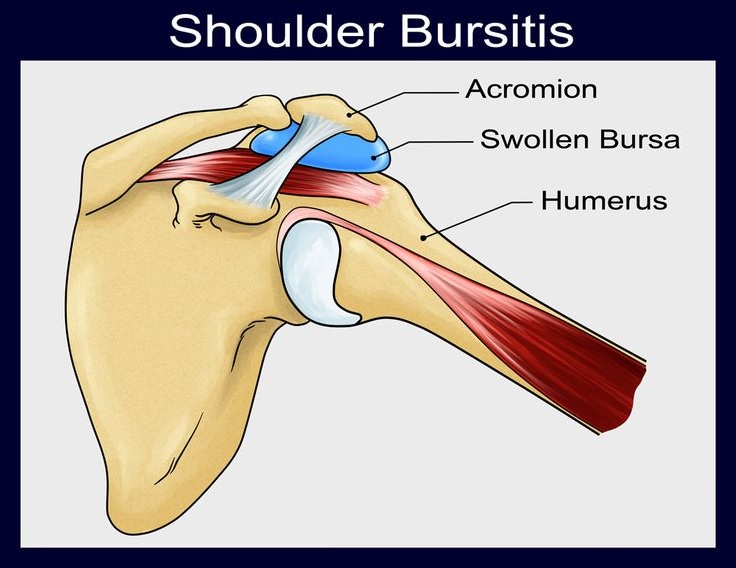
Shoulder Bursitis - What it is, Symptoms, Treatment, Cure


What is shoulder bursitis?

The shoulder bursitis results from an inflammation of the synovial bags (or bursae) that exist around this joint and the tendons of the rotator hood.

It is one of the most frequent causes of pain in this joint. It can be bilateral, reaching both the left shoulder and the right shoulder.

It can be acute or progress to chronic shoulder bursitis, says the orthopaedic in Delhi.

Subacromial bursitis

There are several bursitis that can occur in the shoulder depending on whether one or the other synovial pouch is affected.

The most frequent are those that reach the sub deltoidal subacromial bursa, forming what is commonly called subacromial bursitis.

Shoulder bursitis – symptoms

Shoulder bursitis presents a clinical picture similar to that of tendonitis in the shoulder.

The most characteristic symptoms are the presence of inflammatory pain, located on the antero-lateral face of the shoulder, eventually radiating to the arm and elbow. Its worsening is especially felt with efforts or during the night, making it impossible for the patient to sleep on the affected shoulder.

Also characteristic of shoulder bursitis is the presence of crackling that is palpable, or perceived by the patient, when mobilizing the joint, explains the orthopaedic doctor in Delhi.

Shoulder bursitis – causes

The most frequent causes of shoulder bursitis are trauma and repeated efforts, such as those that occur with certain work activities (painters or plasterers, warehouse replenishers, etc.) or with the practice of certain sports, such as weight training, swimming or others practiced with the arm above the head (“overhead sports”), says the sports injury specialist in west Delhi.

Certain rheumatic diseases (such as, for example, rheumatoid arthritis, gout, lupus, psoriatic arthritis) also often develop with bursitis (namely subacromial).

Shoulder bursitis – diagnosis

The diagnosis of this pathology is made, essentially, through a careful clinical examination, carried out by your specialist shoulder orthopaedic doctor in Delhi.

The exams to be performed later may be an x-ray that helps to exclude other causes of shoulder pain (such as osteoarthritis or calcifying tendinitis) and an ultrasound of the shoulder that easily visualizes the fluid in the subacromial bursa (caused by the inflammatory effusion).

The nuclear magnetic resonance (NMR) of the shoulder is, however, one that has a better diagnostic accuracy, obtaining high-resolution images of the surrounding muscle-tendon structures and eluding any intra-articular pathology, states the orthopaedic in West Delhi.

Is shoulder bursitis curable?

Shoulder bursitis is curable. Next, learn how to treat shoulder bursitis.

Shoulder bursitis – treatment

The medication or remedy most commonly used to treat shoulder bursitis is anti-inflammatory. The most common are ibuprofen, diclofenac, naproxen, among others. Its use is aimed at relieving pain and decreasing the inflammation that is always present in these conditions. Its application can be topical in the form of creams (or gels), or impregnated dressings that will make a prolonged release of these agents in the affected area.

The patient can also take these drugs in oral form (in capsules or diluted), taking care to do so after meals or preceded by taking gastric protectors, in order to avoid the aggression of the gastric or duodenal mucous membranes, states the orthopaedic doctor in Dwarka.

The physiotherapy is essential to maintain joint function and help control pain. A well-oriented physiotherapeutic treatment, with adequate exercises, can prevent the installation of a marked stiffness in the shoulder, usually called adhesive or retractable capsulitis (frozen shoulder or “frozen shoulder”). This would oblige to prolong the treatment for its complete resolution.

In cases of very acute and incapacitating pain, or reluctant to any other non-invasive treatment, infiltration or local injection of corticosteroids diluted with local anesthetic, may be a therapeutic weapon to be used by your orthopaedic doctor in Dwarka to resolve the condition. Your institution should be judicious and always consider the pros and risks of its application.

Shoulder bursitis – surgery

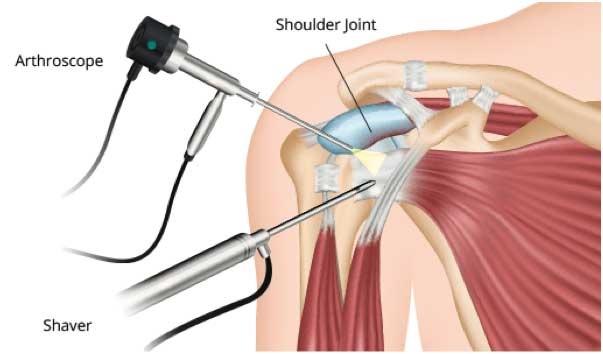
Shoulder arthroscopy in Delhi (surgery) is reserved for cases that are resistant to medical treatment or that simultaneously present other pathologies, such as rotator cuff tears or osteoarthritis, says the orthopaedic surgeon in Delhi.

This operation is performed using mini-invasive techniques that allow an excellent articular visualization, performing a minimal surgical aggression and thus providing a well-tolerated postoperative period and an easier and faster rehabilitation.

It should be carried out in specialized orthopaedic clinic in Delhi and by experienced orthopaedic surgeon in Delhi in order to obtain the best results.

Wednesday, 10 February 2021

What is an Arthroscopy and its Advantages?


Arthroscopy in Delhi is a minimally invasive surgery, which is used for the diagnosis and treatment of the joints. It is performed by introducing an arthroscope connected to a high-resolution camera, which allows the joint to be viewed in its entirety through a high-definition monitor.

This technique has many benefits and is increasingly being used to treat joint diseases. It allows a more complete view of the joint, through minimal incisions, which are made under local anesthesia. As it is a minimally invasive intervention, it offers good results and the patient’s recovery times are reduced, states the orthopaedic in Delhi.

Types of arthroscopy

It could be said that there are no different types of arthroscopy in Delhi, since the technique used is always the same, however, the differences arise depending on the joint operated.

The most common arthroscopies are the following:

  • Shoulder
  • Elbow
  • Doll
  • Hip
  • Knee
  • Ankle

When is an arthroscopic operation performed?

Arthroscopy in Delhi is a technique that is carried out to visualize the inside of the patient’s joint. In many cases, it is carried out as a diagnostic test, for example, to take samples or perform biopsies of the area, in case of tumors or intra-articular lesions.

However, in other cases, it is carried out for the treatment of joint injury, for example, to remove cysts, perform intra-articular washes or remove extra-articular bodies that can cause inflammation of the patient’s joint. It is important that you are always advised by a specialist orthopaedic doctor in Delhi.

How is the arthroscopic operation?

Arthroscopic intervention is usually carried out using local anesthesia at orthopaedic clinic in Delhi, that is, localized anesthesia in the area to be intervened. First, a minimal incision is made by the orthopaedic surgeon in Delhi, through which the arthroscope will be inserted.

Once the incision has been made, cartilaginous cleaning is carried out, that is, debridements are corrected in order to provide stability to the cartilage. In many cases, a treatment with plasma rich in platelets or stem cells is applied, which help the regeneration of the intervened tissues, explains the orthopaedic in Delhi.

Advantages of arthroscopy

Arthroscopic interventions have many advantages over traditional surgeries. First of all, it is a minimally invasive technique, the incisions are of a minimum size, which makes the risk of suffering infections considerably lower.

In addition, the patient recovers quickly and the time it takes to be able to return to their normal activities is reduced, says the orthopaedic in Delhi.

How is recovery after arthroscopy?

As explained, the recovery after an arthroscopic operation is fast, since the invasion into the tissues is minimal. Proper care of the incisions is especially important to avoid infections that can affect recovery.

Usually, the patient will start a rehabilitative treatment one month after the operation, with the aim of strengthening and restoring joint mobility. Three or four months after surgery, the patient will be able to recover his usual activities, avoiding activities that imply an impact on the joint, explains the orthopaedic in Delhi.

Rehabilitation after arthroscopy

In arthroscopic interventions, rehabilitation is of particular importance, which can be carried out under the supervision of an orthopaedic doctor in Dwarka or a specialized physiotherapist in Dwarka. The objective is to strengthen the joint, to prevent the injury from developing again, and to regain joint stability.

In this process, it is advisable to carry out sports practices that do not imply a great impact on the joint, for example, walking. In addition, the patient must perform prescribed flexion and extension exercises, which help to regain full mobility of the joint, says the orthopaedic in Dwarka.

As explained, arthroscopy in Dwarka is a technique that can correct injuries that are disabling for the patient and help him regain his quality of life.

When to Visit an Orthopaedic Specialist

  Orthopaedic conditions can significantly affect your daily life. Recognizing when to see a specialist can help prevent further damage and ...