Thursday 19 May 2022

UNICOMPARTMENTAL KNEE PROSTHESIS


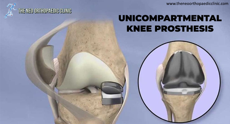
During knee replacement surgery in Delhi, the damaged bone and cartilage are covered with metal and plastic components. In a unicompartmental knee replacement (also called a “partial” knee replacement), only part of the knee is covered. This procedure is an alternative to total knee replacement for patients whose disease is limited to just one area of ​​the knee.

Because partial knee replacement is done through a smaller incision, patients generally spend less time in the hospital and return to normal activities sooner than patients undergoing total knee replacement.

ADVANTAGES OF PARTIAL KNEE REPLACEMENT

Several studies show that most patients who are suitable candidates for the procedure have good results with unicompartmental knee replacement in Delhi.
The advantages of partial knee replacement over total knee replacement include:

  • Faster recovery;
  • Less pain after surgery;
  • Less blood loss;

DISADVANTAGES OF PARTIAL KNEE REPLACEMENT

The disadvantages of partial knee replacement compared to total knee replacement include:

  • Slightly less predictable pain relief;
  • Potential need for more surgery. For example, a total knee replacement in Delhi may be necessary in the future if arthritis develops in the parts of the knee that were not replaced;

SURGERY CANDIDATES

If your osteoarthritis has advanced and non-surgical treatment options are no longer relieving your symptoms, orthopaedic in Dwarka may recommend knee replacement surgery in West Delhi. In order to be a candidate for unicompartmental knee replacement, your arthritis must be limited to one compartment of your knee. In addition, if you have any of the following characteristics, you may not be eligible for the procedure:

  • Inflammatory arthritis;
  • Significant knee stiffness;
  • ligament damage;

With proper patient selection, modern unicompartmental knee replacements have demonstrated excellent mid- and long-term results in younger and older patients.

YOUR SURGERY

A partial knee replacement operation typically lasts between 1 and 2 hours.

Partial knee replacement. There are three basic steps in the procedure:

  • Prepare the bone. Your orthopaedic surgeon in Dwarka will use special saws to remove cartilage from the damaged compartment of your knee;
  • Position the metal implants. The removed cartilage and bone are replaced with metallic coatings that recreate the joint’s surface. These metal pieces are typically held in the bone with cement;
  • Insert a spacer. A plastic insert is placed between the two metal components to create a smooth gliding surface;

COMPLICATIONS

As with any surgical procedure, there are risks involved with a partial knee replacement. Your orthopaedic surgeon in West Delhi will discuss each of the risks with you and take specific steps to help prevent potential complications.

Although rare, the most common risks include:

  • Blood clots. Blood clots in the leg veins are a common complication of knee replacement surgery. Blood clots can form in the deep veins of the legs or pelvis after surgery. Blood anticoagulants such as low molecular weight heparin and aspirin can help prevent this problem. Newer medications, such as rivaroxaban (Xarelto), may also be prescribed by your orthopaedic doctor in Dwarka, depending on your needs;
  • Infection. After surgery, an infection may occur in the skin over the wound or deep into the wound. An infection can happen while you are in the hospital or after you go home. You will be given antibiotics before the start of your surgery and these will be continued for about 24 hours afterwards to prevent infection;
  • Nerve or vessel damage. Although it rarely happens, nerves or blood vessels can be injured or stretched during the procedure;
  • continued pain;
  • Risks of anesthesia;
  • Need for additional surgery;

RECOVERY

Hospital discharge. Patients with partial knee replacement generally experience less postoperative pain, less swelling, and have easier rehabilitation than patients undergoing total knee replacement. In most cases, patients go home 1 to 3 days after the operation. Some patients go home on the day of surgery.

Weight support. You will begin putting weight on your knee immediately after surgery. You may need a walker, or crutches for the first few days or weeks until you feel comfortable enough to walk without assistance.

Rehabilitation exercise. A physiotherapist in Dwarka will give you exercises to help maintain your range of motion and restore your strength.

Tuesday 17 May 2022

Patellar Instability or Patellar Dislocation


What is Patellar Instability or Patellar Dislocation?

The patella, formerly known as the kneecap, is the front bone of the knee, responsible for transmitting the strength of the thigh muscles. In some situations, this bone can move out of its normal location, called patellar instability or patellar dislocation, explains the orthopaedic in Delhi.

If the patellar dislocation occurred for the first time, it is called a primal patellar dislocation. From the second episode, it is called recurrent patellar dislocation.

What are the symptoms of patellar instability?

When a patellar dislocation occurs, there is severe pain and an inability to mobilize the knee. It is possible to see and feel that the patella has moved out of place.

Most of the time, the patella comes back into place on its own almost immediately. Rarely, a doctor needs to put it in place with a knee extension maneuver.

After a patellar dislocation, there may be a feeling of insecurity with the knee, even without the patella clearly moving out of place. This sensation is called a patellar seizure. It is a very uncomfortable symptom, which can interfere with normal activities, says the orthopaedic in Dwarka.

How and why does patella dislocation occur?

Patellar dislocation can occur from trauma, such as a blow or twist to the knee, or without trauma, in a common movement of the joint.

Some people have knee features that favor patellar dislocation. Among the main ones are:

  • High patella
    • The patellar tendon is longer, which makes the patella rest on the knee in a higher position, decreasing the bony socket at the beginning of knee flexion.
  • Increased patellar tilt
  • Femoral trochlea dysplasia
    • The groove on the femur where the patella fits can be shallower than normal, completely flat, or even convex.
  • Increase of the “Q” angle
    • “Q” angle is formed by the direction of traction of the thigh musculature and the direction of traction of the patellar tendon.
  • Patients with valgus knees (knees in, or in “X”)
  • Ligament laxity is also more predisposed.

What is the medial patellofemoral ligament?

The medial patellofemoral ligament is the structure that prevents dislocation of the patella. When the patella is displaced, it is injured or loosened.

When the patellar dislocation is treated without surgery, what is expected is that this ligament will heal. In the surgical treatment of patellar dislocation, reconstruction of the medial patellofemoral ligament is performed in most cases, explains the orthopaedic surgeon in Delhi.

Learn more about patellar dislocation treatments below.

How is patellar instability diagnosed?

The diagnosis of patellar instability is made through a careful assessment of the patient’s clinical history and physical examination, complemented with imaging tests.

The main test to be evaluated is magnetic resonance imaging, which shows indirect signs of dislocation, injury to the medial patellofemoral ligament, and the anatomical changes that favor instability. In addition, MRI is essential to look for cartilage lesions. Other tests, such as radiographs in special positions and computed tomography, are useful for evaluating the shape of the knee and predisposing factors, states the orthopaedic in west Delhi.

How is patellar instability treated without surgery? In what situation is he indicated?

In the case of a patient with an episode of patellar dislocation, both non-surgical and surgical treatment are possible. The decision for one or the other must be individualized, after a detailed discussion between the patient and the orthopaedic surgeon in Dwarka.

Non-surgical treatment involves immobilization for a period, followed by rehabilitation focused on exercises to strengthen and control the thigh and hip muscles. The goal of successful non-surgical treatment is the absence of new episodes of dislocation and patellar apprehension, the feeling of discomfort or buckling caused by instability, explains the orthopaedic surgeon in Dwarka.

In which cases is surgery indicated?

Situations that indicate treatment with patellar dislocation surgery are:

  • Recurrent episodes of dislocation (recurrent patellar dislocation)
  • Association with cartilage injuries
  • Symptoms of apprehension getting in the way of normal activities

Patients with a single episode of dislocation, although they can be treated without surgery, can also opt for surgical treatment. Surgery has the advantage of a lower chance of re-displacement or seizure symptoms for activities, says the orthopaedic surgeon in west Delhi.

What are patellar instability surgeries like?

According to the orthopaedic surgeon in Delhi, there are several procedures available for patellar instability, which are chosen according to the characteristics of each patient, and there may be a combination of procedures. This concept of individualized treatment is known as à la carte treatment, influenced by the French school.

These are the most common procedures performed for patellar dislocation.

  • Reconstruction of the medial patellofemoral ligament
    This ligament is the main restrictor of patellar dislocation, and its reconstruction is indicated in almost all cases. It is a graft from the patient’s own tendon to remake the ligament.
  • Lateral release (or release)
    Release of structures that hold the patella on the side or outside, when there is excess tension. Can be done openly or arthroscopically (video surgery)
  • Tibial tuberosity osteotomy
    A cut is made in the tibial bone to reposition the point where the patellar tendon attaches. This transfer allows for patellar realignment or patellar height correction.
  • Trochleoplasty
    Correction of the shape of the femoral trochlea, the groove where the patella rests on the femur.
  • Treatment of cartilage injuries
    When cartilage injuries also exist, these may also need specific treatment.

How is the postoperative period and rehabilitation?

Postoperative care depends on the technique used. They usually involve a period of support with crutches and a knee brace. However, from the beginning, it is already allowed to put the foot on the floor and remove the immobilizer to move the knee in most situations. Rehabilitation includes restoring knee mobility and restoring strength and control of the musculature of the thigh, hip, and trunk.

Thursday 5 May 2022

What are the symptoms of hip bursitis?


Bursitis in the hip is an inflammation in a hip pouch, called a “bursa”. It is a relatively common problem and is known to cause pain and generate various discomforts for the patient. It is part of the great trochanter pain syndrome and is associated with hip tendinitis as well. Being treated by the best orthopaedic in Dwarka, this disease unfortunately leaves many patients with their movements reduced, as well as prevents the hip from being flexed, for example. It can be caused by several factors, bursitis is uncomfortable, but with the correct diagnosis can be easily treated.

Hip expert: knowing what bursitis is important to fight it!

Bursitis is a problem that can appear in both sedentary people and sportsmen and happens because of repetitive movements and overloads in the hip joint. Knowing your symptoms, causes and treatment is essential to get rid of these limiting pains and avoid the causes of bursitis. With the correct follow-up with the orthopaedic in Dwarka, this nightmare can have your days numbered. Learn more about bursitis and stay tuned for symptoms.

Pain

This is the main symptom of bursitis and is present in almost all cases. The most common complaint of patients with this problem is pain in the lateral face of the hip. This pain worsens when sleeping on the inflamed hip. The sensation is burning pain and usually worsens if the patient does not change position.

Discomfort for movement and pain to walk, get up and sit are also quite common, not to mention that staying for long periods of time in the same position can cause worsening of the pain as well. The pain of bursitis is characterized by radiating to the thighs, often confusing the patient, who thinks that the problem may be in another location.

Limitation

Slow movements, not being able to walk as before, having pain when tying shoes and cannot squat are the main signs of the limitation of bursitis. Because it is an inflammation of the bursas that are located on the lateral face of the joint, bursitis can actually cause problems in movements and in turn limit the daily activities of the patient.

Swelling

Bursitis can cause local swelling, which in turn can be felt manually by the patient. The location besides being palpable, may also be with increased temperature.

Pain to the touch

Touch pain also happens in bursitis. Often when touching the swollen region, the patient may feel pain. This happens because of the whole inflammatory process and also by the daily activities of the patient.

Hip bursitis is usually very painful and limiting. The patient is usually very concerned about the symptoms, but as a correct treatment, the resolution prognosis is usually excellent.

Causes of hip bursitis

The causes of the appearance of hip bursitis are varied:

  • Repetitive overload injury. Running, climbing stairs, cycling, or standing for long periods of time.
  • hip injury
  • Spine diseases.
  • Differences in the length of the legs.
  • Rheumatoid arthritis.
  • Hip surgeries or hip replacement in Delhi that can irritate the bursa and cause bursitis.
  • Bone spurs or calcium deposits that can develop in the tendons and irritate the bursa.

How long does hip bursitis last?

The duration of trochanteric bursitis cannot be determined in advance. In some cases, such as trauma, it can last for a short period of time, but sometimes if one is neglected it can become chronic.

When the pain subsides, begin to move slowly back to normal movement. At this time, it is important to have the support of a physiotherapist in Dwarka who will indicate the exercises to be performed to improve movement in the area.

With these simple actions, the bursitis will subside. But if it doesn’t, corticosteroid injections can be used, growth factors can be introduced to repair damaged tissues, or even combined with ozone therapy. The last option is surgery, if the previous methods are not effective.

How to treat hip bursitis?

In the treatment of hip bursitis, rest is essential, at least while the pain remains in its acute phase. Let your hip rest as long as possible. To do this, it may be convenient to use a cushion with an anatomical design and memory.

This cushion distributes the weight, avoiding excessive pressure and achieving adequate pelvic stability. With it you will be able to rest the hip and relieve the pain. And when you sleep, it is best to do it on your back or on the opposite side of the affected area, placing some pillows between your knees.

In addition, it is convenient to apply cold to the hip to reduce pain and inflammation. The orthopaedic in West Delhi will also recommend taking non-steroidal anti-inflammatory drugs.

Without a doubt, the best treatment for hip bursitis is prevention. It is necessary to avoid that it appears for the first time or that new episodes arise. That’s why it’s important:

  • Avoid activities that overload the hip, such as being on your feet for a long time.
  • Lose weight if you are overweight or obese, to take pressure off the area.
  • Sit on cushions, to avoid excessive pressure in the area.
  • Do warm-up and stretching exercises before and after doing any physical exercise.
  • Avoid muscle atrophy with the performance of specific exercises.
  • Wear a comfortable shoe.
  • Correct bad posture and do not do activities that can cause pain in the area.

And above all, follow the advice of orthopaedic doctor in Dwarka to try to prevent hip bursitis from becoming chronic. Thus, you will avoid pain and the muscles in the area from atrophying.

The Revolution of Robotic Knee Replacement Surgery

Introduction Robotic knee replacement surgery has emerged as a groundbreaking advancement in the field of medical science. This innovative...