Friday 27 March 2020

MENISCUS DAMAGE

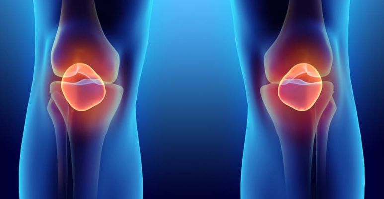
The meniscus is a semi-circular structure inside the knee located between the joint areas of the femur and tibia.

The meniscus distributes the load and protects the articular cartilage, and also partly gives stability to the knee while walking. Inside the knee are two menisci - internal and external. The external meniscus is more mobile; therefore, it is injured much less often. A meniscus injury is a fairly common occurrence, which is most common among physically active citizens.

Despite this, in a small number of cases, menisci are damaged during various degenerative processes inside the knee, for example, osteoarthritis, explains orthopaedic in Delhi.

HOW IS MY KNEE ARRANGED?
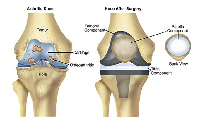
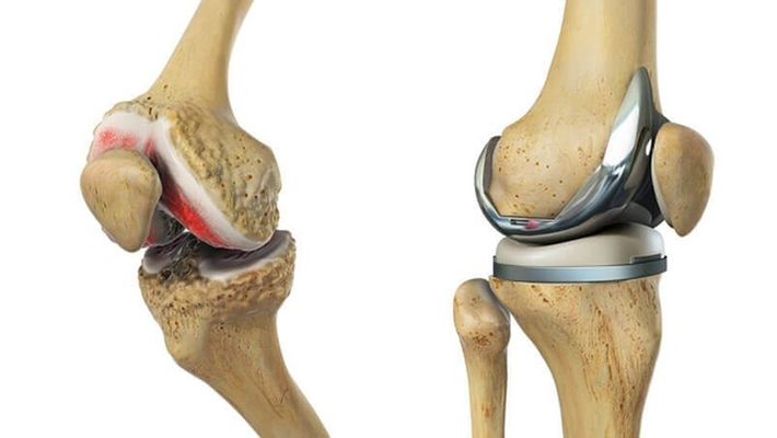
The knee joint consists of three bones (femur, tibia, and patella), connected rigidly together with the help of ligaments that stabilize the joint. The articulating surfaces of the bones inside the joint are lined with a smooth protective tissue called articular cartilage, it allows the bones to glide relative to each other.

With arthritis and arthrosis, as well as damage to the menisci and ligaments, cartilage is significantly damaged.

Ligaments are dense structures of connective tissue that hold bone to bone and stabilize the knee. Inside the knee joint are two main ligaments. An anterior cruciate ligament (ACL) and posterior cruciate ligament (PCL). They cross crosswise in the center of the joint, from which they got this name. The other two main ligaments are actually located outside the knee joint, on the outside and inside of the knee. They act to stabilize the lateral movement of the knee.

The ligament on the inside of the knee is called the medial collateral ligament or MCL. The ligament on the outside of the knee is called the lateral collateral ligament or LCL.

The patellar ligament connects the lower pole of the patella with the upper part of the lower leg. The central third of this ligament is sometimes used as a source of graft when restoring a torn anterior cruciate ligament.

The meniscus is a crescent-shaped or C-shaped structure located between the joint areas of the thigh and lower leg. There are two menisci in each knee, one on the inside called the “medial meniscus” and one on the outside called the “lateral meniscus”.

The meniscus is one of the varieties of cartilage. The meniscus can be torn during twisting movements in the knee. Rupture can occur along the inner edge of the meniscus, or, less commonly, along the outer edge. There can also be only a small meniscus gap, for example, a patchwork or a large so-called “handle-watering” gap; it represents a gap along the entire length of the meniscus. Such a gap can block the joint, which means that the leg cannot be straightened to the end. All types of meniscus tears can be successfully treated with arthroscopy in Delhi.

WHAT IS THE MECHANISM OF MENISCUS DAMAGE?
Menisci most often burst during sports, with sharp rotational and rotational-flexion movements in the knee, deep squats, and jumps. A torn meniscus fragment moves into the joint cavity during movements and causes sudden pain and periodic blockages. Meniscus tears are often accompanied by trauma to the cruciate and lateral ligaments of the knee. Due to the peculiarities of meniscus nutrition, they grow to the place of separation extremely rarely, says orthopaedic in West Delhi.

HOW CAN YOU SUSPECT A MENISCUS INJURY?
Common symptoms with a fresh joint injury are inflammation, severe pain, limited mobility in the knee, and fluid buildup in the joint. According to the abatement of the acute period, after a few weeks, the symptoms characteristic of the meniscus rupture come to the fore: local pain at the level of the joint space, “clicks”, “crackling” during movements, muscle hypotrophy, inflammation of the synovial membrane with the formation of effusions, periodic blockages.

HOW TO DIAGNOSE MENISCUS INJURY?
The menisci of the structure are cartilaginous; therefore, they are not visualized on x-rays. The diagnosis is made by a specialist orthopaedic in Dwarka based on complaints, clinical test results, and magnetic resonance imaging data.

WHAT TO DO IMMEDIATELY AFTER AN INJURY?
Immediately after a knee injury, orthopedic in Delhi recommends:
  • immediate limitation of knee loads, moving on crutches;
  • placing a cold compress on the joint, this will reduce pain and swelling, as well as stop bleeding inside the joint. - it is better to use an orthosis or gypsum in a pinch for fixing the knee;
  • giving the limb an elevated position will also help reduce swelling and pain in the knee;
  • take non-steroidal anti-inflammatory drugs.
HOW TO CURE A TORN MENISCUS?
Treatment of meniscus lesions is more often surgical. Using arthroscopy of the meniscus, partial (partial) or less often complete removal is performed. It is also possible to put stitches on the meniscus and fix it to the tear point, but unfortunately, this does not in all cases give the planned result.

With minor injuries of the meniscus, non-surgical treatment is possible. Indications for surgery are large gaps characterized by mechanical symptoms (clicks, crunching, pinching, limitation of mobility), the recurrent formation of fluid in the joint, as well as in cases of unsuccessful non-surgical treatment. It is worth emphasizing that with meniscus tears orthopaedic surgeon in Delhi does not advise to postpone treatment and put up with poorly tolerated pain in the knee.

A torn meniscus and its fragments can irreversibly destroy articular cartilage, up to its erasure to the bone. In modern conditions, meniscus surgery is performed using arthroscopy. Arthroscopic intervention is performed through two punctures up to 0.7 cm long. An arthroscope is connected to one of the arthroscopes through the camera to the monitor, and instruments for manipulating the joint are inserted through the other.

Meniscus arthroscopy is performed in an aqueous medium. With arthroscopy, it is possible to achieve excellent cosmetic results, the method is not very traumatic, accelerated rehabilitation. The arthroscope allows us to the most distant parts of the joint, the examination of which is not always possible with open operations explains orthopaedic in Janakpuri.

Thursday 26 March 2020

Total Knee Arthroplasty

Total-Knee-Arthroplasty

What is:
  • Arthroplasty / total knee replacement in Delhi is a surgical intervention in which the worn-out joint surfaces of the femur, tibia and often the kneecap are replaced, as they are a source of disabling pain. Metallic and high-density polyethylene components are placed in place of damaged and painful joint surfaces.

Surgery objectives:
  • The main objective of placing a total knee prosthesis is to treat the pain caused by arthrosis and, at the same time, to improve joint mobility, increase its functional capacity and return quality of life. In some cases, arthrosis results from a misalignment of the knee in varus or valgus (knees arched or knees together, respectively), and then there is a second objective for knee replacement in Dwarka, which is the correction of this anatomical deformity.
  • The joint mobility to be achieved after the surgery will be between 100 to 130 degrees of flexion and the full extension. However, the main factor that determines the postoperative amplitude is the amplitude before surgery, so a knee with reduced mobility will have more difficulty in reaching the intended amplitude.

Indications:
  • All patients with an indication for total arthroplasty are frankly limited to activities of daily living, suffering from an intense and disabling painful condition, resistant to medical and physiatric therapy and without any other surgical option. The age factor should be evaluated according to the currently available statistical data, which shows that the survival of total knee replacement in Delhi is 90% at 10 and 80% at 20 years old (defined by the prostheses still functioning) respectively. However, the placement of knee prostheses can be proposed at younger ages, in particular clinical situations that have no other medical or surgical alternative, which will return the patient's quality of life. The majority of patients are in the age group above 60 years, with the upper limit today dependent on general clinical condition, with many cases operated after age 80. The surgical risk will always be assessed in the Preoperative Anesthesiology Consultation. Another important factor, regardless of age, is the motivation of the patient and his family, in the face of disability and suffering caused by arthrosis, after an open discussion of the risks and benefits, especially if there are no serious pathologies, which may condition the patient's longevity.
  • The patient with an indication for this surgery has more frequently developed osteoarthritis of idiopathic etiology (a cause is not identified), mechanical due to axis deviation or post-traumatic, due to sequelae of joint fractures.
  • Rheumatic diseases, especially rheumatoid arthritis, represent the second most important group of pathologies, due to their joint involvement.
  • Excessive body weight is not a contraindication, as long as the patient has a general condition compatible with the surgery. The long-term results are slightly lower, as far as surgery is concerned, however, it must be considered that obesity associated with limited mobility is itself a vicious cycle, both due to the medical pathologies that it originates and due to the greater body weight gain.
  • The longevity of the prosthesis mentioned above, motivates us to try whenever possible a more conservative surgical approach in younger patients, defined by age groups below 60 years of age. This approach consists of arthroscopic gestures, osteotomies for axis correction (“straightening crooked legs”) or even physiotherapy associated with medication and infiltrations in the context of regenerative medicine.

Complications / Risks:
  • Performing an arthroplasty implies risks that must be known to the patient. Possible postoperative complications include thromboembolism, which motivates mandatory pharmacological prevention, skin complications, and neurovascular injuries. The infection, often referred to as rejection, has an incidence of less than 1%, but may require new surgery to wash and replace the prosthesis.
  • The failure of arthroplasty may be due to aseptic detachment (loose components in the absence of infection, due to sensitivity to polyethylene wear) and premature wear of the material. In these cases, it also involves performing a new surgery for its revision/replacement, states orthopaedic in Dwarka.

Surgical technique:
  • The surgical technique we use today is called minimally invasive. It consists in the use of smaller skin incisions, but above all in a lesser aggression of the surrounding soft parts (muscles, synovial membrane, ligaments, vessels, and nerves), explains the orthopaedic in Delhi.
  • This approach allows for a less painful postoperative period, less blood loss, less risk of infection and a faster and less painful recovery. Most patients are able, after an initial period with support and load of the operated lower limb, with 2 crutches, to resume a gait without assistants for 1 to 2 months, depending on the condition of the contralateral knee.
  • In performing this surgery, general or loco-regional anesthesia can be used, depending on the patient's and anesthetist's decision, during the Anesthesiology Consultation.
  • The rehabilitation program starts during hospitalization, about 48 hours after surgery, leaving the patient to walk with the help of 2 crutches and able to go up and downstairs.

Postoperative Care:
  • The placement of a knee arthroplasty implies limitations for the practice of sports and should avoid activities with impact. However, walking, golf, gym, swimming, dancing, and cycling are allowed. Patients should promote the longevity of their prosthesis, avoiding overweight, intense physical efforts with impact (running), transporting heavy loads and hyperflexion postures.
  • Any infection that occurs, anywhere in the body, must be treated early and effectively, due to the risk of bacteremia (bacteria circulating in the bloodstream) that can contaminate the knee prosthesis from a distance. For the same reason, antibiotic prevention should also be systematically applied to all dental or endoscopic manipulations, using your attending physician whenever necessary, suggests the orthopaedic in Delhi.

Tuesday 24 March 2020

Hip Arthroplasty

Hip-Arthroplasty

Hip Prosthesis Surgery

The hip replacement in Delhi is an operation performed with a 10-12 cm incision and through which makes resection of the femoral head and the acetabular cartilage to allow for its replacement by an implant (prosthesis) of metal.

The hip prosthesis can be fixed to the bone by applying a special cement (cemented prosthesis), which adheres and hardens within a few minutes, allowing the load to patients immediately after hip replacement surgery in Delhi. It is ideal for older patients, with more osteoporotic bone, or who have worse bone stock due to rheumatism pathology.

Cementless prostheses are applied under pressure (press-fit). They are produced with a rough and porous surface, usually coated with hydroxyapatite, to allow a process of incorporation by bone growth into the interior of your pores, allowing a very firm and lasting fixation.

Generally, the application of a total hip prosthesis does not require general anesthesia, but a locoregional block, with the placement of a catheter to control postoperative pain.

Total Hip Arthroplasty

hip replacement surgery in West Delhi is a surgery that has undergone a major evolution. Its application is possible by mini-invasive techniques, which provide the patient with better and easier recoveries.

Also, in terms of instruments for placing the total hip prosthesis, technical advances have been made in order to make them more precise and versatile with solutions for the different types of hip to be replaced. This evolution was based on a better knowledge of the anatomy and biomechanics of the joint.

Finally, the interfaces between the femoral and acetabular components have also improved. In addition to the classic metal-polyethylene, it is possible to apply ceramic components that show wear rates and production of smaller particles, and therefore, allowing greater durability of the implants.

Partial Hip Arthroplasty

In some situations, in which the acetabulum is spared and a very elderly patient has suffered a fracture of the femoral neck, it is possible that the Orthopaedic in Delhi may choose to perform a partial hip arthroplasty, replacing only the femoral head and thus making a procedure simpler and less invasive for the patient.

This saves operational time and blood loss, which can be important in patients who are very weak and whose general condition needs to be optimized.

Hip Arthroplasty - Risks, Complications

Hip replacement in West Delhi is not without risks and complications.

First of all, there are complications that can occur in any joint replacement surgery in Delhi, regardless of the joint. These are hemorrhages, infections or venous thrombosis.

Then there are the specific complications of hip arthroplasty. At an early stage, during and immediately after surgery, vascular injuries (including injuries to the sciatic nerve), dysmetria and instability may occur, with the occurrence of dislocation of the prosthesis. In a later stage, periprosthetic fractures or even detachments of the prosthesis may occur.

Currently, the technical solutions available to the orthopaedic surgeon in Delhi are increasingly varied and versatile to solve these situations, namely with the use of the so-called hip arthroplasty revision surgery.

Hip Arthroplasty - Recovery

The recovery after performing total hip arthroplasty, is increasingly rapid and simple for the patient, since the surgery is less invasive, more effective control of pain and more accelerated rehabilitation protocols.

The recovery time is thus substantially reduced. Lift and load on the operated limb are allowed on the first day after surgery, with discharge to hospital generally occurring between the 3rd and 4th day. The stitches are removed at 15 days in the outpatient consultation and the patient usually dispenses with the use of external gait supports (Canadian), at around 3-4 weeks.

In our clinical practice, on the 1st postoperative day, the patient undergoing a total hip prosthesis begins a physiotherapy regimen, with a bi-daily visit by a specialized Physiotherapist in Delhi. Immediate mobilization prevents the occurrence of deep venous thrombosis, prevents muscle atrophy and facilitates lifting.

Physiotherapy starts immediately with exercises taught during hospitalization, namely gait training and up and downstairs.

After the first 2 weeks after a total hip replacement in Delhi, the exercises are specifically aimed at strengthening the muscles involved in walking, especially the glutes or swimmers (namely the middle).

Hydrotherapy is also recommended at a more advanced stage of recovery, in order to improve joint tone and proprioceptivity, as well as increase the patient's general well-being and confidence.

Hip Prosthesis Surgery - Price

The price of a hip surgery can vary according to the type of surgical intervention and the type of prosthesis placed. The amount may also fluctuate if the patient has any health insurance and associated conditions.

Only the orthopedic in Delhi can determine how much the operation costs, through a thorough analysis in the consultation.

Friday 20 March 2020

Symptoms and Treatment of Knee Dislocation

Knee-Dislocation

Overview

In order to better understand what dislocation refers to at one knee or even at both knees, we need to know some information about the anatomical structure of the knee.

The patella is most often prone to dislocation because it is part of the knee joint and has the role of facilitating the connections between the thigh muscles and those of the tibia.

Due to its up-down movement when we flex and extend the leg at the time of trauma, the patella slides out of the patellar capsule.

Another lesser-known name for the patella is the kneecap, being a small sesamoid bone with a round shape, but flattened anterior-posteriorly with the anterior face in a triangular shape, which can palpate under the skin, thus forming the anterior knee relief.

This time of dislocation is more frequent in women, but also in men, especially young people, around the age of 20 years.

Dislocation can occur either as a result of a minor trauma or accidentally due to a wrong movement during a sport or other contexts.

Dislocation of the knee should not be confused with the sprain of the knee. The dislocation also involves affecting the other parts of the knee joint, while the sprain, being a milder condition, produces only a strain of the ligaments or even their rupture in extreme cases, but without the bones coming out of the joint.

And the pain is different in the two situations. In the case of knee dislocation, the pain is very intense and mobilization with foot support is impossible, while in the case of a knee sprain, the pain is bearable, and mobilization can be achieved.

Causes

The causes that lead to dislocation of the patella are given by:
  • trauma to the knee;
  • falls from the height;
  • sports accidents after which the knee can be twisted;
  • femoral-patellar dysplasia;
  • patellar chondropathy;
  • malposition of the patella given by the lateral insertion of the patella tendon;
  • genus valgum (the shape of the legs in X);
  • positioning the patella at a higher level, which can be a congenital cause;
  • violent contact with another adversary by abruptly hitting the knee.
It should be mentioned that when the dislocation of the patella occurs accidentally, the situation can be a serious one because blood vessels, nerves can be damaged, there is the possibility of meniscus rupture or even the joint capsule may suffer.

If we talk about the event that the disruption occurs in athletes either by hitting the opponents or by falling to the knee, the consequences are less severe and, most of the time, do not accompany additional injuries.

Signs and symptoms
  • the primary sign is the vivid and deafening pain accompanied by the characteristic "cracking" (cracking) noise;
  • followed by rapid swelling of the area;
  • deformation of the affected region;
  • the impossibility of mobilizing the lower limb.
The diagnosis is most often made during the clinical examination when the orthopaedic in Delhi observes the signs and symptoms mentioned above. To see the exact location of the patella, it can perform an x-ray.

Another paraclinical examination recommended in special cases is the MRI, in order to detect possible ligament ruptures, cartilage or synovial membrane damage, components that cannot be visualized on plain radiography.

In the event that the patella replenishment occurs spontaneously either by yourself or by another person until you reach the orthopaedic in Dwarka, you should not overload the joint or force it or even delay the visit of orthopaedic in West Delhi because there may be small neurovascular lesions that may accentuate in the following hours or days.

It should also be mentioned that a timely treatment is 80% likely to have a solid and stable joint in the future, while lack or delay of treatment for various reasons reduces the chances of success by 20% and increase the risk of gonarthrosis.

Treatment

The initiation of orthopedic treatment is mainly based on pain relief and prevention of complications.
The orthopedic reduction translated by repositioning the joint is intentional besides the relief of pain. This repositioning will be performed under general anesthesia, in case of surgery.

Another relatively modern surgical treatment is the arthroscopic one, with numerous advantages for the patient, starting from the reduced incisions in size and to the shorter recovery time.

Arthroscopy in Delhi can reconstruct some injured ligaments during dislocation and reduce joint stiffness.

Infiltration treatment is renowned for the benefits of platelet-enriched plasma. This plasma will be introduced at the level of the affected knee, in order to recover rapidly from the collateral lesions.

We must not omit the immobilization of the knee, which is done by putting the plaster (gypsum) of the knee for a period of 3 weeks to 2 months.

Kinetotherapy also plays an essential role in the recovery of patients after a knee dislocation.

The recovery program is established in collaboration with the kinetotherapist and the orthopaedic surgeon in Delhi, depending on the treatment applied and can be extended for up to 1 year.

Depending on the evolution of the patient, the degree of difficulty of the exercises can be intensified, but with the limitation of the flexion and extension movements of the knee in order not to overload the joint.

Physiotherapy in Delhi is also recommended, which aims to reduce local inflammation of the affected knee.

Prevention

There are no special measures to be taken because, for the most part, accidents are not scheduled and can happen anytime, anywhere.
However, going to the gym or playing sports helps strengthen the muscles, thus reducing the risk of knee dislocation.

Monday 16 March 2020

Everything You Need To Know About Shoulder Subluxation

Shoulder subluxation refers to the partial dislocation of the shoulder joint. This happens when the upper part of the humerus (humeral head) partially exits the glenoid cavity of the shoulder blade. In a complete dislocation, the humerus is completely removed from the joint cavity.

The shoulder is the most mobile joint of the human body. This joint contains a series of bones, ligaments, and muscles that work together to maintain a stable shoulder. However, due to the high mobility, the shoulder is very susceptible to dislocation.

Thus, a subluxation is often the result of trauma or injury. Also, another cause can be the stroke, because it can weaken the muscles in that area.

Clinical picture
A subluxation may be more difficult to identify than a complete dislocation. However, in some cases, the partially dislocated humerus may be visible under the skin.

Also, the bone can easily feel palpable and can cause discomfort and pain. The most common symptoms of subluxation include:
  • The possibility to observe the deformation of the shoulder;
  • Pain;
  • Inflation;
  • Sensation of numbness or tingling in the arm (paresthesia);
  • Functional impotence;
  • Sensation of shaking or interrupting movements during the activity.

Causes
Because the shoulder can move in several directions, it can move forward, backward or downward. This is also true for subluxations.
When the dislocation is partial, the joint capsule may be affected, including rupture, which can lead to different complications.
Typically, strong hits or falls can cause a shoulder dislocation. Also, the extreme rotation of the arm can cause the humerus to be pulled out of the glenoid cavity.
Once a shoulder has been dislocated, the joint may become unstable and prone to future dislocations or subluxations.
Shoulder subluxation is mostly caused by:
  • Trauma - subluxation can result from accidents or injuries that affect the joints or other structures that provide stability to the shoulder. The most common examples are falls and road accidents.
  • Sports accidents - contact sports, but not only these, can often cause shoulder subluxation. Gymnastics and skiing are other sports with a high degree of risk, due to frequent falls.
  • Stroke - this neurological condition can cause muscle weakness. Thus, the destabilization of the shoulder joint can be complicated by a subluxation.
A small study reported that 80% of stroke participants also suffered a shoulder subluxation.
Young men, as well as groups of extremely physically active people, have the highest risk of shoulder subluxation.

Treatment options
Proper diagnosis is essential for adopting the best treatment option. Thus, very good diagnostic methods consist of ultrasonography of the shoulder joint or radiography.
The treatment aims to reposition the humerus back to the original place and ensure that it stays in position. Treatment options include:
- Closed reduction - this intervention involves easy handling of the bone back into position. In most cases, severe pain disappears almost immediately after reducing maneuvers.
- Surgical intervention - it is recommended by orthopaedic in Delhi especially when the dislocations are repeated. This is also the preferred treatment when the nerves, blood vessels or ligaments of the shoulder have been damaged.
- Orthosis - some people may need to wear orthotics for a few days or weeks to prevent the shoulder bones from moving. The length of time this device should be worn depends on the severity of the trauma.
- Medicines - after surgery, but before, the orthopaedic surgeon in Delhi may recommend a muscle relaxant and an anti-inflammatory drug. In this way, pain and swelling can be reduced.
- Rehabilitation - After surgery, the orthopaedic in Dwarka may indicate rehabilitation programs. Their purpose is to restore the range of motion, endurance, and shoulder joint.
The physiotherapist in Delhi may recommend certain exercises that can help you recover faster. These should be done correctly and under supervision, as in certain circumstances, they may aggravate the symptoms. Also, massage and ice applications may be required.

Recovery and potential complications
When an injured person has a subluxation without major injury to the nerves and tissues, the simple repositioning of the humerus should rapidly improve the patient's condition.
However, if a rest period is not respected and normal activities returned, it is possible to dislocate the shoulder later.
After surgery for a subluxated shoulder, the orthopaedic in West Delhi will recommend, most of the time, that the person in question should wear shoulder support for several weeks.
The gradual introduction of physiotherapy can help a person regain his or her power and movements. However, it is not indicated the intense movements of the shoulder and the activities not recommended by the orthopaedic in Janakpuri. Thus, subsequent complications can be prevented.
The shoulder contains strong tissues, muscles, and ligaments, all helping to maintain the humerus in its place. However, these structures can be injured, complicating subluxation.

Thus, the most common complications of a shoulder subluxation include:
- Affecting blood vessels and shoulder nerves;
- Temporary or permanent loss of movement and flexibility;
- Instability at the shoulder level, followed by recurrent subluxations;
- Other lesions in the tissues of the affected shoulder.

Differential diagnosis
Shoulder subluxation presents some common symptoms with other similar lesions. For this reason, there may be errors in the diagnosis. A subluxation can be confused with:
  • Biceps tendonitis - the condition refers to inflammation of the biceps tendon. Due to the position of this tendon, its damage can usually lead to shoulder pain.
  • Clavicular injuries - fractures or traumas of the clavicle can cause pain in the shoulder, but also in the hindrance of movements at this level.
  • Rotator cuff injuries - this structure covers much of the shoulder joint capsule. His injuries can cause symptoms like those of the shoulder subluxation. Also, minor injuries can heal on their own, while severe injuries may require surgery.
  • Shoulder dislocation - dislocation involves the complete separation of the humerus from the joint cavity. The symptoms are like those of subluxation, and the correct differentiation of these conditions can only be made by an orthopedic in Delhi.
  • “Swimmer's Shoulder” - Shoulder pain in professional swimmers is often referred to as this.
Because swimming requires a high level of shoulder flexibility and a wide range of movements, swimmers are often likely to have an increased risk of a shoulder injury.

Perspective
When a person requests medical attention quickly and receives a correct diagnosis, shoulder subluxation is completely treatable.
However, when no surgery is required, it may take several months for the person to resume normal activity.
The recovery time varies depending on the extent of the subluxation and whether or not the affected person has undergone surgery.
Also, following a subluxation, the person concerned must avoid intense activity, to prevent relapse and to speed up recovery.

Friday 13 March 2020

Everything We Need To Know About Finger Sprain


The sprain of the finger occurs when a bone from its formation (a phalanx) moves out of the joint. Injuries resulting from sports activities, falls or accidents can cause dislocation of the fingers.

Dislocation or sprained ness of a finger can be extremely painful, but not life-threatening. However, it is very important to receive medical support from orthopaedic in Dwarka as quickly as possible.

Symptoms of finger sprain
The symptoms of finger sprain are not numerous, but they successfully manage to indicate the condition. Thus, the affected finger changes its shape and looks crooked, but it is also particularly painful.

Other signs that may indicate finger sprain include:
- numbness or tingling;
- bruising or discoloration in the skin;
- functional impotence.

What are the causes of finger sprain
Normally, the thumb contains two phalanges, and the rest of the fingers contain three. Thus, there are two joints in the case of the thumb and three joints for the other fingers.

Ligaments are strips of fibrous tissue that help keep the joints integrated. Dislocation occurs when a significant force overcomes the opposite resistance of ligaments and causes the bones out of the joint.
Injuries suffered from sports are the most common causes of finger sprains. Thus, it is estimated that over 50% of the injuries found in the sport target fingers. Sports with the highest rates of injuries to the hands include football, gymnastics, basketball and wrestling.

Other possible causes that may lead to a sprain of the finger are:
- exaggerated extension of the finger;
- applying a strong blow to the tip of the fingers;
- falls;
- the existence of other health conditions that can weaken the joints and ligaments.

First measures in case of finger sprain
People who suspect their finger is sprained should seek medical attention from orthopaedic in Delhi immediately. To relieve inflammation and immediate pain, ice can be applied, but with great care, since the injured finger should not be moved.

The affected person or the people around you should not attempt to place the bone back in the joint. The only people who can do this are those who have medical training in this field.

Trying to re-encase the bone by unprepared people can seriously aggravate the situation, causing additional pain and inflammation. In addition, moving their own bones back to the original place comes with a very high risk of permanently harming the surrounding structures (tendons, ligaments, nerves, blood vessels, joint cartilage).

Diagnosis of finger sprain
After arrival at the orthopaedic in Janakpuri, it will examine the affected finger. The orthopaedic in West Delhi should also be informed when the trauma occurred and what were the circumstances of the production.

Further investigations may be requested after primary evaluation to confirm the diagnosis or to assess the severity of the lesion. The necessary imaging investigations are:
- X-rays are used to observe the internal structures of the body, in this case the bones. X-rays can help confirm the diagnosis of luxuryness, but also check if there are certain associated fractures;
- magnetic resonance imaging – MRI scanning uses strong magnetic fields and radio waves. With their help, detailed images of the tissues inside the body are obtained. In most cases, this method is required if significant damage to the tissue adjacent to the lysed joint is suspected.

Treatment options
Treatment options vary depending on the location and severity of the sprain. Thus, the methods of treatment are:

Reduction of sprain
The first step in treating a dislocated finger is to carefully handle the bone and relocate it into the joint. The procedure is known as a discount.

Before performing local anesthetics can be used to relieve pain, and after the procedure has been completed, x-rays can be performed to check its effectiveness.

Immobilisation
After the reduction, the patient is recommended to wear an immobilization soften. It protects and secures the wounded finger as it heals.
Also, wearing the band stops the patient from moving the finger and prevents further dislocation and injury.
Wearing softening by patients is necessary for about a few weeks.
However, immobilization for prolonged periods can cause permanent rigidity and reduced finger mobility.

Fixing
Depending on the type or severity of injury, some people may suffer alongside sprains even bone fractures. Fractures are defined as staples or interruptions in a bone.
Bone fractures also require reduction and immobilization, but fixation may also be needed. Fastening is carried out with K brooches, these are thin metal rods that surgeons attach to stabilize bone fragments.

Surgery
Dislocated fingers involving torn ligaments and complex bone fractures may require surgical procedures known as open cuts. Like other treatments, surgeries are aimed at reducing, stabilizing and restoring the mobility of the affected finger, without damaging the surrounding structures.

Recovery

According to orthopaedic surgeon in Delhi, finger luxations usually heal in about 4-6 weeks.
The factors affecting the recovery time are as follows:
- severity and location of dislocation;
- the existence of ligament damage or tendons;
- the existence of bone fractures;
- the need for surgery.

As a result of reduction and immobilization, certain patients may require physical or occupational therapy. The physiotherapist in Delhi can help and instruct the patient how to speed up his healing and how to expand his range of movements.

There are also certain tips to be followed at home:
- maintenance of skins, but also clean and dry steas;
- maintaining the affected finger at a higher level than the heart to reduce inflammation;
- avoiding the request of the affected finger;
- application of cold compresses and ice packets to reduce pain and inflammation;
- administration of medicines against pain and inflammation, such as ibuprofen or acetaminophen;
- performing exercise just as recommended by the specialist.

It should be known that a finger recovered from a sprain presents a high risk of further injury in the future. Thus, caution is recommended when physical activities are carried out, even wearing protective equipment and avoiding risk sports.

Conclusions
Dislocation of a finger can cause panic and extremely much pain. However, it should be known that finger sprain is not life-threatening, but requires prompt medical assistance.
No attempt should be made to reduce home sprain by the patient or his close friends. In this way complications can occur, and healing can be hampered, sometimes there is a risk of permanent injuries.

Visit to the orthopedic in Delhi is mandatory, it can correctly assess the located to provide adequate treatment. After treatment, healing usually lasts a few weeks. However, if the sprain is accompanied by bone fractures or damage to adjacent tissues, the recovery time may be prolonged.

Wednesday 11 March 2020

Chronic L5 / S1 Back Pain is not "NORMAL"

L5-S1-Disc

80% of the population have at least one round of debilitating low back pain at least once in a lifetime. Between 10 and 15 percent of the population has problems in the lower back that recur repeatedly throughout their adolescent and adult lives. Many people have back problems that originate from the lumbosacral joint, which is where the lower fifth lumbar disc meets the first sacral disc of the spine, L5 / S1, explains the orthopaedic in Delhi.

In plain language, this is the part of the lower back where the branches of the spine change to the pelvis and legs. In reality, this structure is not a single "joint." It includes a disc between the lumbar and sacral spine, and two facet joints (also known as zygophysical joints) that guide the movement of this part of the spine.

Many things can go wrong with this part of the spine. The disc may herniate. When this happens, the internal contents of the disc seep and touch the adjacent nerve. This condition is known as a lumbar herniated disc, explains the orthopaedic surgeon in Delhi.

Or the disc itself can degenerate. This condition is known as degenerative lumbar disc disease, or DDL. Another common possibility is that the facet joints fracture so that they cannot hold the lumbar disc in place. It glides over the sacrum in a condition known as L5-S1 isthmic spondylolisthesis. This part of the spine can also be damaged by stenosis, narrowing, or degenerative arthritis.

The spinal cord does not extend down into the lumbar spine. There is no danger of paralysis when this part of the spine is damaged. On the other hand, there is no end to the pain that can be generated by damage to this part of the spine. However, that doesn't mean that just because your lower back is killing you, you don't have a potentially serious condition. Sometimes you should not try to get pain care at home, and you need to see an orthopaedic in Dwarka right away:
  • Sudden bowel or bladder problems, either the inability to "go" or the inability to hold it, which appear suddenly, can be a sign of an urgent medical problem.
  • Severe back pain along with stomach pain from being unable to get up is also a potential sign of an emergency medical condition.
  • Weakness in the legs with problems in bowel or bladder control is also a signal to see a doctor right away.

However, chronic lower back pain can be treated in several ways at home:
  • Take pain relief as prescribed by the orthopaedic in Dwarka. If you need more pain relief, see the same doctor to request a change to your prescription. Avoid taking too much Tylenol (acetaminophen) to avoid liver damage.
  • Deep point massage (acupressure) is helpful in controlling pain but will not cure your back problems.
  • Movement-style acupuncture treatment (which receives acupuncture during exercise) is used in some Asian treatment centres.
  • Approximately two months of constant home exercise is required to restore strength and range of motion in the lower back. You cannot do the exercises again and hope to improve. Consistency is key. Your doctor can give you the specific exercises you need to do.
  • You may need a session with a physical therapist or coach.
  • Your orthopaedic in Delhi may prescribe a biofeedback machine to tell you when you are moving in a way that supports the recovery of your back.
  • Device-based therapy, using an exercise machine like the David Spine Concept system, is successful approximately 75 percent of the time. These machines are found in well-equipped gyms. You should not try to work out your own routine on ordinary gym equipment unless you are a physical therapist or coach.
  • If you are exercising well enough to exercise and have not been given specific exercises to strengthen your back, exercise with a Swiss ball and rubber bands instead of machines, at least at first. Exercise lightly with minimal effort until you are sure you can exercise without causing pain.
  • If you play contact sports, going back to the game before an injury anywhere else on your body increases your risk of lower back injury. The problem is you can't respond to a hit, kick, or tackle fast enough to avoid stress on your spine. A first injury tends to lead to a second or third, says the sports injury specialist in west Delhi.

If there's good news about low back pain and all the conditions discussed here, it's that they usually go away eventually even without treatment, if you can avoid injuring your back again. It can take weeks, months, or even a year or two, but almost all of them eventually get better.

Saturday 7 March 2020

Knee Replacement Surgery

Knee-Prosthesis

The purpose of knee replacement in Delhi is to replace the knee, or the part of the knee with a prosthesis, which is damaged by osteoarthritis. In some cases, it is an implant that will replace the articular surfaces (cartilage) of the tibia, the femur and the patella.

Who is it for?

In general, since implanting a prosthesis is a definitive surgical act, it can be suggested by the orthopaedic in Delhi when the patient's pain cannot be calmed with any other medical or surgical therapeutic method that is normally used in case of osteoarthritis.

Generally, this operation is performed in patients over 65 years of age, in which osteoarthritis affects two or more internal, external and anterior knee compartments. But some young patients may be the subject of an exception, each case is different, and only the doctor can judge the need and benefit of such a surgery.

The surgery

The knee replacement in Delhi is an important surgery. It requires a preparation that includes a complete clinical and radiological balance. If possible, it must be done in an establishment specializing in prosthetic surgery. Normally, the duration of hospitalization exceeds one week. This operation requires general or epidural anaesthesia.

In general, this decision will depend on your general condition and the type of operation that requires the implantation of your prosthesis. The duration of the operation can vary between 50 minutes and 2 hours. Generally, after the operation, it is convenient to stay between 15 and 30 days in a re-education centre, explains the best knee surgeon in Delhi.

The days after the operation and the re-education

You can start slowly walking towards the fourth day, with some crutches that are necessary at the beginning, and then can be abandoned (usually, after a month and a half). The re-education advisable or even essential. The help of the physiotherapist and the surgeon will allow to recover the amplitude of the movements and facilitates the resumption of the march. Normally, the operated person can return to work after 2 and a half months or 3 months.

However, after a surgery of this magnitude, the patient should periodically visit his family doctor (or General practitioners, Rheumatologists, Orthopedists, Traumatologists, Internists, Physiatrists, Rehabilitation Medicine). For this, the consultations to the orthopaedic surgeon in Delhi must also be regular: between 2 and 3 months after the surgery, another after 6 months and another one year later. Next, an annual check is recommended.

The results

Pain and mobility

The knee replacement in Dwarka is often effective in alleviating pain. It suppresses between 80 and 95% of the pain of osteoarthritis. The improvement of mobility depends on the type of prosthesis used, but also on the condition of the patient before the operation. However, generally, there is clear progress.

The scar

Normally, the scar is located on the vertical axis of the leg, in front of the kneecap.

The type of prosthesis

There are different types of prostheses that differ depending on:
  • The part of the knee replaced: partial or complete prosthesis (tri-compartmental prostheses)
  • The mode of fixation on the knee joint
  • The knee stabilization medium used
  • The mobility of the prosthesis components: fixed or mobile plateau.

Tri-compartmental prostheses

This type of prosthesis is the most frequent. They can be used in most osteoarthritis, even those that affect a single compartment of the knee. In addition, there are several models and they are used according to the state of deformation of the knee and allow the preservation or not of the cruciate ligaments. The choice of one of these prostheses depends on the state of your knee. Since each case is different, your orthopaedic in west Delhi is the best person to guide you in choosing a model. Tri-compartmental prosthetic models vary depending on 3 factors:
  • Knee fixation mode: with cement or without cement
  • The stabilization system: The different models allow the two crossed ligaments to be well preserved, one or none.
  • The mobility of the fixed or mobile plateau.

The patellofemoral prosthesis

Osteoarthritis of the joint between the patella and the femur. This type of prosthesis is used exceptionally.

Partial or single compartment prostheses

Generally, this type of prosthesis allows an improvement in the mobility of the knee and hip. In addition to an almost complete disappearance of pain. Unlike the other two types of prostheses, these allow only the worn part of the joint to be replaced. However, this type of prosthesis is only used when only one of the knee compartments is affected: the internal, external femoro-tibial compartment or, exceptionally, the joint between the patella and the femur.

In addition, its implantation can only be advised in a patient whose knee deformation is unimportant and has normal cruciate ligaments. Your orthopaedic in Delhi is the best counsellor to explain whether you can benefit from this type of prosthesis.

Tuesday 3 March 2020

Hip Replacement Surgery: Risks and Complications

Hip Replacement

The hip joint consists of the pelvic bone, which has a receptacle to receive the end (ball) of the femur. When the cartilage or joint bones are diseased or severely damaged, a surgical procedure called total hip replacement can be done to remove the diseased ball and replace them with prosthetic materials or hip implants, explains the orthopaedic in Delhi.

The goals of hip replacement surgery in Delhi are to improve joint function and relieve chronic symptoms of severe pain. It is usually done due to the progressive worsening of arthritis in the hip, which is often due to degenerative arthritis or osteoarthritis. This condition is usually associated with aging, congenital anomaly or trauma to the joint.

Other conditions that can lead to total hip replacement in Delhi include rheumatoid arthritis, fractures of the hip joint and death (necrosis) of the hip bone. Necrosis can be caused by hip fracture, chronic drug use, alcoholism and some diseases (such as lupus).

Hip replacement surgery in Delhi is an important surgical procedure that involves some risks and complications. Apart from the usual complications that are associated with most surgeries in the immediate postoperative period (such as bleeding, infection, fever, pain, nerve injury and blood clots), the surgical procedure also carries some long-term risks and complications. It could happen months or years after a patient is discharged from the hospital. These can lead to implant failure, which may be due to several causes, such as:
  • Loosening of implants in the absence of infection. This is a common problem, which can be caused by several factors such as the type of implant chosen, the surgical technique and the amount of force applied to the joint by the patient.
  • Bone fracture
  • Component fracture
  • Late infection
  • Late dislocation
  • Joint stiffness
  • Abrasive forces in the union that lead to the production of debris
  • Bone loss (osteolysis), which occurs when the body reacts to eliminate waste when it is recognized as foreign particles. This is the most frequent long-term hip replacement complication, causing almost 75% of all hip implant failures. Severe joint pain can result from debris production and osteolysis.

Orthopaedic surgeon in Delhi usually refer their patients to a rehabilitation team after surgery to help them with physical and occupational therapy. Patients are instructed to observe some precautions for several weeks to avoid dislocation of their new hip. These include:
  • Do not bend the hip more than 90 degrees
  • Don't cross your legs
  • Do not turn the affected leg or too far

Total rehabilitation usually takes at least six months. Patients are also placed on an exercise program to follow after going home, even while still having physical therapy to help speed their recovery. Weight control is also necessary to reduce stress on the hips. However, one must remain active to maintain flexibility, strength and endurance. Activities may include walking, swimming, dancing and riding a stationary bicycle. The most exhausting activities, such as tennis or jogging are not advisable by the orthopaedic in Dwarka.

Most hip joint implants last 10 to 20 years without loosening. This can depend on factors such as the patient's lifestyle, the amount of stress that is given to a joint, the weight of the patient and how well the healing process goes, says the orthopaedic in Delhi.

The Revolution of Robotic Knee Replacement Surgery

Introduction Robotic knee replacement surgery has emerged as a groundbreaking advancement in the field of medical science. This innovative...