Showing posts with label orthopaedic surgeon in Delhi. Show all posts

Thursday, 11 April 2024

The Revolution of Robotic Knee Replacement Surgery

Introduction

Robotic knee replacement surgery has emerged as a groundbreaking advancement in the field of medical science. This innovative procedure combines the precision of robotics with the expertise of orthopedic surgeons to provide patients with improved outcomes and faster recovery times. In this article, we will explore the benefits of robotic knee replacement surgery and how it is revolutionizing the field of orthopedics.

What is Robotic Knee Replacement Surgery?

Robotic knee replacement surgery, also known as robotic-assisted knee surgery, is a minimally invasive procedure that utilizes advanced robotic technology to assist orthopedic surgeons during knee replacement surgeries. This procedure involves the use of a robotic arm, which is controlled by the surgeon, to perform the surgery with enhanced precision and accuracy.


The Benefits of Robotic Knee Replacement Surgery

1. Enhanced Precision

One of the key advantages of robotic knee replacement surgery is the enhanced precision it offers. The robotic arm used in this procedure allows for more accurate placement and alignment of the knee implant. This precision helps to optimize the function and stability of the knee joint, leading to improved outcomes for patients.

2. Customized Treatment

Every patient is unique, and their knee replacement surgery should reflect that. Robotic knee replacement surgery enables surgeons to create a personalized treatment plan based on the patient's specific anatomy. By using advanced imaging technology, the surgeon can develop a 3D model of the patient's knee, which helps in determining the optimal size and placement of the implant. This customized approach ensures a better fit and alignment, leading to improved patient satisfaction.

3. Minimally Invasive Procedure

Robotic knee replacement surgery is a minimally invasive procedure, meaning it requires smaller incisions compared to traditional open surgery. The smaller incisions result in less trauma to the surrounding tissues, reduced blood loss, and a faster recovery time for the patient. Additionally, the use of robotics allows for a more precise and targeted approach, minimizing the risk of damage to healthy tissues.

4. Faster Recovery

Due to the minimally invasive nature of robotic knee replacement surgery, patients typically experience a faster recovery compared to traditional surgery. The smaller incisions result in less pain and scarring, allowing patients to regain mobility and return to their normal activities sooner. This accelerated recovery time is particularly beneficial for individuals who lead active lifestyles or have demanding physical jobs.

Is Robotic Knee Replacement Surgery Right for You?

Robotic knee replacement surgery is a highly advanced procedure that offers numerous benefits. However, it may not be suitable for everyone. Your orthopedic surgeon in Delhi will evaluate your specific condition and determine if you are a good candidate for robotic-assisted knee surgery. Factors such as the severity of your knee condition, your overall health, and any previous surgeries will be taken into consideration.

Conclusion

Robotic knee replacement surgery in Delhi is a revolutionary advancement in the field of medical science. By combining the precision of robotics with the expertise of orthopedic surgeons, this procedure offers enhanced precision, customized treatment, and faster recovery times for patients. While not suitable for everyone, robotic knee replacement surgery provides a promising option for individuals seeking improved outcomes and a quicker return to their active lifestyles. Consult with your orthopedic in Dwarka to determine if robotic-assisted knee surgery is right for you. 

Wednesday, 17 August 2022

Achilles tendon rupture - treatment, surgery


What is Achilles tendon rupture?

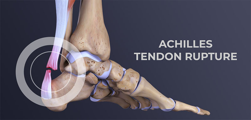
An Achilles tendon rupture or rupture occurs when the tendon “ruptures or tears” leading to separation or discontinuity in the tissues that make up the tendon.

In terms of anatomy, the Achilles tendon, a sort of fibrous “ribbon” or “cord” that connects muscle to bone, is the largest and strongest tendon in the body that connects the calcaneus (heel bone) to the medial gastrocnemius muscles., lateral gastrocnemius and soleus muscles (popularly known as calf muscles or “leg belly” muscles). When the muscle contracts, it “pulls” the tendon, which in turn moves the foot.

The rupture often reflects the previous existence of tendinosis phenomena caused by sports microtraumas or degeneration (aging) of the tendon. In most cases, the tear occurs at the bottom of the tendon (just above the heel), but it can occur anywhere along the tendon. In the presence of a healthy tendon, the lesion can appear in the bone or muscle.

Total ruptures, partial ruptures

We can generically classify Achilles tendon ruptures into:

  • Total rupture – in the total rupture of the Achilles tendon, the tissues break completely, that is, the tendon is completely “separated”.
  • Partial rupture – in a partial rupture of the Achilles tendon, the tendon does not completely rupture, only an incomplete rupture occurs. Partial tears can vary greatly in severity according to the extent of the injury.

A total rupture is more frequent than a partial rupture. As a rule, a total rupture is a more serious injury when compared to a partial rupture and with more exacerbated symptoms. A partial tear can also present with pain that can range from moderate to severe and, if not recognized, it can quickly progress to a total tear.

See more information on treatments to better understand the different therapeutic approaches according to the extent of the lesion.

Achilles tendon rupture – causes

The Achilles tendon can lose elasticity and become “weak and thin” with age and lack of use. Then, it becomes prone to injury or breakage.

Rupture is more common in people with pre-existing Achilles tendonitis (tendon inflammation). Repetitive tendinitis and the consequent calcifications are a risk factor for tendon rupture.

Certain diseases (such as arthritis and diabetes) and medications (such as corticosteroids, for example) can also increase the risk of rupture.

Rupture occurs most often in the middle-aged male athlete. The injury usually occurs during recreational sports that require impacts with the ground, running, jumping, etc. Most of the time, these are football, athletics, tennis, basketball, among others. Injury can happen in these situations as a result of traumatic dorsiflexion when the muscle is strongly contracted causing it to tear.

Breaks can, however, also occur in everyday activities. For example, when falling from a significant height, when entering a pothole abruptly, traffic accidents, etc.

Achilles tendon rupture – symptoms

The signs and symptoms of Achilles tendon rupture are:

  • Sudden, severe pain may be felt in the “back” of the ankle or the “tummy of the leg”, often described as “being hit by a rock or shot” or “like someone stepping on the back of the ankle”;
  • A loud clicking sound can be heard;
  • A discontinuity (“gap”) or depression (void) can be felt and seen in the tendon above the calcaneus (heel bone);
  • Inability to stand on tiptoe on the affected side;
  • Initial pain, swelling (swelling) and stiffness may be followed by bruising and weakness (not being able to land on the foot, walking).

Achilles tendon rupture – diagnosis

The diagnosis is made by the orthopaedic doctor in Delhi after collecting the clinical history, performing the physical examination and some complementary means of diagnosis (MCDT).Bottom of Form

A simple test is to “stretch” the “calf or calf muscles” while lying on your stomach (Thompson test). In the impossibility of being able to elevate the foot, there is, most likely, a rupture in the tendon. This test isolates the connection between the “calf muscles” and the tendon and eliminates other tendons that may still allow poor movement.

The orthopaedic doctor in Dwarka may order the following tests to confirm the diagnosis and to know in greater detail the location and degree of severity of the lesion:

  • Plain radiography (XR) – not being a very useful exam, it can identify a bone fragment avulsion of the calcaneus;
  • Ultrasound or ultrasound – Ultrasound of the leg and thigh can help assess the possibility of deep vein thrombosis and can also be used to rule out a Baker’s cyst (or cyst). Ultrasound can identify Achilles tendon rupture or signs of inflammation (tendinitis or tendinosis);
  • Magnetic Resonance Imaging (MRI) – MRI is extremely sensitive for diagnosis and allows you to determine if there is still a tendon in continuity. It allows other diagnoses such as tendinitis, tendinosis and bursitis.

Pain in the “back of the heel” is not always due to an Achilles tendon rupture. In the differential diagnosis, tendinitis (inflammation of the Achilles) and bursitis (inflammation of the bursae) should be considered, among the most frequent pathologies that cause pain in the Achilles region.

Achilles tendon rupture – treatment

The objective of the treatment is to restore the function of the tendon, for this, it is necessary that the tissues that make up the tendon heal “united” with each other. In this way, it will be possible for the patient to return to the same level of activity before the injury. Regaining Achilles tendon function after an injury is critical to making walking possible.

Treatment reflects a balance between tendon protection and initial movement. Protection is necessary to allow time for healing and to prevent further injury. Moving the foot and ankle is necessary to prevent stiffness and loss of muscle strength.

We can divide treatment options into surgical and non-surgical. Conservative (non-surgical) treatment consists of a set of therapeutic attitudes aimed at healing the tendon and restoring its function without resorting to any type of surgical intervention.

The choice between surgical and non-surgical treatment can be controversial in some cases. Both surgical and non-surgical treatment will require an initial period of approximately six weeks of immobilization. For most patients, both treatment options have good functional results.

NON-SURGICAL TREATMENT

Nonsurgical treatment is often used for non-athletes or for people with a low general level of physical activity who will not benefit from surgery. In the elderly and people with clinical complications, conservative (non-surgical) treatment should also be considered as a first option.

Initially, a cast below the knee is performed with the foot in equinus (foot in marked plantar flexion, “down”). Although it is not routine, it is possible to perform an MRI to verify that the tops of the tendon are in contact. The cast is changed, at intervals of two to four weeks, to slowly stretch the tendon back to its normal length. This treatment usually takes 8 to 12 weeks. During this period, global strengthening and flexibility exercises are taught.

SURGERY (SURGICAL TREATMENT)

Surgery on Achilles tendon rupture is often indicated in healthy and active people who want to resume activities such as walking, running, cycling, etc. Even those who are less active may be candidates for surgical repair of the tendon. The decision to operate should be discussed with your orthopaedic surgeon in Delhi.

Surgery should not be performed if there is an active infection or damaged skin at or around the Achilles tendon rupture site. In addition, some diseases or lifestyle habits, such as diabetes, smoking habits, sedentary lifestyle, steroid use and inability to follow instructions after the operation, may be a contraindication for surgery.

Surgical intervention for an Achilles tendon rupture is usually performed on an outpatient basis. This means that the patient is operated on and goes home the same day.

We can identify two distinct surgical approaches:

The first is to perform the intervention percutaneously, allowing to perform a minimally invasive surgery, through small incisions. A kind of needles with attached sutures are passed, allowing the Achilles tendon to be sutured.

The second approach is the open approach (traditional method, where the surgeon makes an open incision to access the tendon). This starts with an incision made in the back of the leg, just above the calcaneus (heel bone). After the best orthopaedic in Dwarka finds the two ends of the torn tendon, these ends are sutured. The incision is subsequently closed.

The surgical technique will be previously determined by the orthopaedic in West Delhi, depending on the type and location of the rupture, among other factors. In the postoperative period, regardless of the surgical technique chosen, the patient is immobilized with an equinus foot.

Despite being a safe surgery, some complications can arise, such as risks associated with anesthesia, infection, damage to nerves and blood vessels and bleeding or blood clots, among others. A new rupture can also occur (recurrence).

Recovery after surgery

After the surgery, the patient is placed with a splint or a “plaster boot” from the foot to the knee. Usually, the patient cannot walk or put weight on the involved leg. Crutches, a walker or a wheelchair are used to allow the patient to remain mobile for the first few times. Patients are encouraged to keep the operated leg elevated above the level of the heart to decrease swelling (swelling) and pain.

Patients are usually seen in the office two weeks after surgery. The splint or cast is removed, and the surgical incision is evaluated. Stitches are usually removed at this point if necessary. After two to six weeks, depending on postoperative protocol and orthopedic in Dwarka preference, patients may be allowed to begin performing some force. For this, a “hiking boot” can be used. Ankle movement is often allowed and encouraged.

After six weeks, full-body strength is generally allowed. Physiotherapy in Dwarka will need to be started and is intended to restore ankle range of motion. Strengthening of the “calf muscles” and Achilles is gradually allowed as the tendon heals. It is usually possible to resume full activity after six months. Recovery time after surgery can extend up to a year, until the patient can achieve full rehabilitation.

Even in cases where surgery is performed, the above-mentioned therapeutic attitudes are included in the rehabilitation plan. See more information on conservative treatment.

Tuesday, 17 May 2022

Patellar Instability or Patellar Dislocation


What is Patellar Instability or Patellar Dislocation?

The patella, formerly known as the kneecap, is the front bone of the knee, responsible for transmitting the strength of the thigh muscles. In some situations, this bone can move out of its normal location, called patellar instability or patellar dislocation, explains the orthopaedic in Delhi.

If the patellar dislocation occurred for the first time, it is called a primal patellar dislocation. From the second episode, it is called recurrent patellar dislocation.

What are the symptoms of patellar instability?

When a patellar dislocation occurs, there is severe pain and an inability to mobilize the knee. It is possible to see and feel that the patella has moved out of place.

Most of the time, the patella comes back into place on its own almost immediately. Rarely, a doctor needs to put it in place with a knee extension maneuver.

After a patellar dislocation, there may be a feeling of insecurity with the knee, even without the patella clearly moving out of place. This sensation is called a patellar seizure. It is a very uncomfortable symptom, which can interfere with normal activities, says the orthopaedic in Dwarka.

How and why does patella dislocation occur?

Patellar dislocation can occur from trauma, such as a blow or twist to the knee, or without trauma, in a common movement of the joint.

Some people have knee features that favor patellar dislocation. Among the main ones are:

  • High patella
    • The patellar tendon is longer, which makes the patella rest on the knee in a higher position, decreasing the bony socket at the beginning of knee flexion.
  • Increased patellar tilt
  • Femoral trochlea dysplasia
    • The groove on the femur where the patella fits can be shallower than normal, completely flat, or even convex.
  • Increase of the “Q” angle
    • “Q” angle is formed by the direction of traction of the thigh musculature and the direction of traction of the patellar tendon.
  • Patients with valgus knees (knees in, or in “X”)
  • Ligament laxity is also more predisposed.

What is the medial patellofemoral ligament?

The medial patellofemoral ligament is the structure that prevents dislocation of the patella. When the patella is displaced, it is injured or loosened.

When the patellar dislocation is treated without surgery, what is expected is that this ligament will heal. In the surgical treatment of patellar dislocation, reconstruction of the medial patellofemoral ligament is performed in most cases, explains the orthopaedic surgeon in Delhi.

Learn more about patellar dislocation treatments below.

How is patellar instability diagnosed?

The diagnosis of patellar instability is made through a careful assessment of the patient’s clinical history and physical examination, complemented with imaging tests.

The main test to be evaluated is magnetic resonance imaging, which shows indirect signs of dislocation, injury to the medial patellofemoral ligament, and the anatomical changes that favor instability. In addition, MRI is essential to look for cartilage lesions. Other tests, such as radiographs in special positions and computed tomography, are useful for evaluating the shape of the knee and predisposing factors, states the orthopaedic in west Delhi.

How is patellar instability treated without surgery? In what situation is he indicated?

In the case of a patient with an episode of patellar dislocation, both non-surgical and surgical treatment are possible. The decision for one or the other must be individualized, after a detailed discussion between the patient and the orthopaedic surgeon in Dwarka.

Non-surgical treatment involves immobilization for a period, followed by rehabilitation focused on exercises to strengthen and control the thigh and hip muscles. The goal of successful non-surgical treatment is the absence of new episodes of dislocation and patellar apprehension, the feeling of discomfort or buckling caused by instability, explains the orthopaedic surgeon in Dwarka.

In which cases is surgery indicated?

Situations that indicate treatment with patellar dislocation surgery are:

  • Recurrent episodes of dislocation (recurrent patellar dislocation)
  • Association with cartilage injuries
  • Symptoms of apprehension getting in the way of normal activities

Patients with a single episode of dislocation, although they can be treated without surgery, can also opt for surgical treatment. Surgery has the advantage of a lower chance of re-displacement or seizure symptoms for activities, says the orthopaedic surgeon in west Delhi.

What are patellar instability surgeries like?

According to the orthopaedic surgeon in Delhi, there are several procedures available for patellar instability, which are chosen according to the characteristics of each patient, and there may be a combination of procedures. This concept of individualized treatment is known as Ã  la carte treatment, influenced by the French school.

These are the most common procedures performed for patellar dislocation.

  • Reconstruction of the medial patellofemoral ligament
    This ligament is the main restrictor of patellar dislocation, and its reconstruction is indicated in almost all cases. It is a graft from the patient’s own tendon to remake the ligament.
  • Lateral release (or release)
    Release of structures that hold the patella on the side or outside, when there is excess tension. Can be done openly or arthroscopically (video surgery)
  • Tibial tuberosity osteotomy
    A cut is made in the tibial bone to reposition the point where the patellar tendon attaches. This transfer allows for patellar realignment or patellar height correction.
  • Trochleoplasty
    Correction of the shape of the femoral trochlea, the groove where the patella rests on the femur.
  • Treatment of cartilage injuries
    When cartilage injuries also exist, these may also need specific treatment.

How is the postoperative period and rehabilitation?

Postoperative care depends on the technique used. They usually involve a period of support with crutches and a knee brace. However, from the beginning, it is already allowed to put the foot on the floor and remove the immobilizer to move the knee in most situations. Rehabilitation includes restoring knee mobility and restoring strength and control of the musculature of the thigh, hip, and trunk.

Wednesday, 20 April 2022

Everything About Knee Replacement

THE KNEE JOINT

Joints are the areas where bones meet, and movement occurs. The knee joint is made up of the femur above and the tibia below. The two bones are separated by cartilage that acts as a cushion and allows movement.

REASONS FOR SURGERY

The reasons for total knee replacement surgery in Delhi are: severe pain, loss of mobility, or deformity of the knee. Symptoms may be due to osteoarthritis, rheumatoid arthritis, or trauma among others.

Osteoarthritis, commonly called "wear and tear," is the most common cause for a total knee replacement.

ABOUT KNEE REPLACEMENT

The knee joint is made up of the ends of the thigh bone (femur) and the shin bone (tibia). These bones normally slide over each other with ease because they are covered by soft cartilage. If an injury damages the cartilage or is worn away by arthritis, for example, it can make the joint ache or stiff.

Generally, a new knee joint improves mobility and decreases pain, although your new knee will not be able to bend as much as a normal knee joint.

Depending on the condition of your knee joint, they will replace part or all of your knee joint. A total knee replacement is more common.

Artificial knee pieces can be made of metal and / or plastic, and a knee replacement can last up to 20 years.

WHAT ARE THE ALTERNATIVES?

Knee replacement surgery in Delhi is generally recommended only if nonsurgical treatments, such as physical therapy and exercise, taking medication, or using physical support devices such as a cane, no longer help decrease pain or improve mobility.

Alternative surgical procedures include arthroscopy in Delhi (if the arthritis is not very severe) or osteotomy (in which the leg bones are cut and put back). You may have already had these procedures before your knee replacement.

The surgeon will explain your options.

PREPARING FOR A KNEE REPLACEMENT

The orthopaedic surgeon in Delhi will explain how to prepare for the operation. For example, if you smoke, they will ask you to stop smoking, as this increases your risk of chest and wound infection, which can delay your recovery.

Typically, you must stay in the hospital for about five days, and the surgery is performed under general anesthesia. This means that you will be asleep during the operation. Otherwise, if you prefer, the surgery can be performed under epidural or spinal anesthesia. This type of anesthesia completely numbs from the waist down, and you will remain awake during the operation.

If you are going to have general anesthesia, you will be asked to fast. This means that you should not eat or drink, normally, for about six hours before general anesthesia. However, it is important to follow the instructions of your anesthetist.

In the hospital, the nurse can check your heart rate and blood pressure and do a urine test.
Your surgeon will explain to you what will happen before, during, and after the procedure, and any pain you may have. This is your opportunity to understand what will happen, and it may be helpful to prepare questions about the risks, benefits, and other alternatives to the procedure. This will help you stay informed so that you can give your consent if you are asked to sign a consent form to carry out the procedure.

You may be asked to wear compression stockings on your unaffected leg to prevent blood clots from forming in your veins (deep vein thrombosis, DVT). You may need an injection of a blood-thinning medicine called heparin in addition to, or instead of wearing, compression stockings.

WHAT HAPPENS DURING A KNEE REPLACEMENT?

Generally, a knee replacement in Delhi takes about two hours.

The orthopaedic in Delhi will make a single cut (10 to 30 cm long) in the front of your knee. You will push the kneecap to the side to reach the knee joint. The surgeon will remove the worn or damaged surfaces from the end of the femur and the top of the tibia. Typically, he will remove the anterior cruciate ligament and may remove the posterior cruciate ligament. For support, the best orthopaedic in Dwarka will not remove the collateral ligaments. It will shape the surfaces of the femur and tibia to fit the artificial knee joint and then fit the new joint over both bones.

Sometimes the back of the kneecap is replaced with a piece of plastic. This is known as patella lining.
After placing the new joint, the surgeon will close the wound with stitches or clips and cover it with a bandage. The surgeon will place a tight bandage on your knee to help minimize swelling.

WHAT SHOULD I EXPECT AFTER


You will need to rest until the anesthesia wears off. After epidural anesthesia, you may not be able to feel or move your legs for several hours.

You may need pain relievers to ease any discomfort when the anesthesia wears off.

You may have an intermittent compression pump attached to special pads on your lower legs for the first day or so. By inflating the cushions, the pump encourages healthy blood circulation and helps prevent a DVT. You can also have a compression stocking on your unaffected leg. This helps maintain circulation.

A physiotherapist in Dwarka (a movement and mobility specialist) will visit you daily to guide you through exercises that will help you recover.

You will stay in the hospital until you can walk safely with the help of a cane or crutch. When you can go home, you will need to ask someone to drive you.

Before you go home, the nurse will give you recommendations for caring for your knee and a date for your follow-up appointment.

How long it takes for the sutures to disappear will depend on the type used in the surgery. However, for this procedure they usually go away in about six weeks. Nonabsorbable sutures and clips are removed 10-14 days after surgery.

RECOVERING FROM KNEE REPLACEMENT SURGERY

If necessary, you can take an over-the-counter pain reliever, for example acetaminophen or ibuprofen. Follow the instructions in the patient information leaflet that comes with your medicine, and if you have questions, ask your pharmacist.

Physical therapy exercises are an indispensable part of your recovery, so it is essential that you continue to do them for at least two months.

You will be able to move around your house and go up and down stairs. For a few weeks, some everyday activities, such as shopping, will be difficult for you to do. You may need to use a cane or crutches for about six weeks.

You may be asked to wear compression stockings at home for several weeks.

When resting, raise your leg and support your knee to help prevent leg and ankle swelling.

Depending on the type of work you do, you may be able to go back to work after six to 12 weeks.

Follow your surgeon's recommendations for driving. You should not drive until you are sure that you can brake in an emergency without discomfort.

WHAT ARE THE RISKS?

Knee replacement surgery in West Delhi is a common and generally safe procedure. However, in order to make an informed decision and consent, you must be aware of the possible side effects and risk of complications associated with this procedure.

Side effects

These are the unwanted, though mostly temporary, effects of successful treatment; for example, feeling dizzy as a result of general anesthesia.

Your knee will hurt and be swollen for up to six months.

You will have a scar on the front of your knee. You may not have sensation in the skin around the scar. This may be permanent, but it should get better in two years.

Complications

Complications are problems that occur during or after the operation. Most of the people are not affected. Possible complications from any operation include unexpected reactions to anesthesia, excessive bleeding, or clot formation, usually in a vein in the leg (DVT).

Complications specific to knee replacement are rare, but include:

  • Wound or joint infection Antibiotics are given during and after surgery to prevent this complication.
  • Unstable joint. The knee joint may loosen and may require surgery to correct it.
  • Damage to blood vessels or nerves. It is usually mild and temporary.
  • Scar tissue. Scar tissue formation can limit movement. You will likely need another surgery to correct it.


Sunday, 17 April 2022

Hip arthroscopy in athletes: eliminate exercise pain

Every athlete who suffers from hip pain usually faces a very similar journey: when the first symptoms appear, they start conservative treatment with anti-inflammatory drugs and painkillers. When the pain improves a little, they try to return to activity. After a little while, they are in pain again.

But when drug treatment no longer has any effect and the pain starts to impair sports practice or even the performance of simple routine activities, what can amateur and high-performance athletes do?

The first step is to seek an orthopedic in Delhi specializing in the hip, who will be able to perform a thorough evaluation. Only after that, treatment will start based on the cause of the injury, that is, try to treat the problem at its root instead of just treating the pain.

The imbalance of strong structures around the hip joint, such as tendons, ligaments and bursae are the main cause of hip pain in those who practice sport. This happens because the hip makes an important connection between the legs and the trunk. Any slight alteration can generate instability and injury.

While many athletes have no indication for surgery, for others, performing a surgical procedure, such as hip arthroscopy in Delhi, is the best option to restore quality of life, sports performance and prevent an injury from becoming chronic, or even progress to cartilage wear.

Hip arthroscopy for athletes

Problems such as hip injuries are closely linked to the practice of sports such as running, cycling, volleyball, tennis, football, dance, artistic gymnastics and other sports.

In the recent past, a large number of young people with hip pain during physical activity did not have a correct diagnosis and some cases progressed to wear and tear. The orthopaedic doctor in Delhi acted as a spectator of the worsening of the joint injury, without being able to interfere with the inevitable.

Today, it is known that many of these young athletes had a disease caused by a change in the fit of the frame, called femoroacetabular impingement.

Athletes with this type of hip injury are the main beneficiaries of hip arthroscopy surgery in Delhi.

Considering the limitations in sports performance imposed by the hip injury, the procedure is a great treatment option for femoroacetabular impingement with labrum injury, as it is capable of correcting the anatomy of the joint socket.

Today, there are several studies that demonstrate that the rate of return to sport after arthroscopy is above 90%. The rehabilitation phase after hip arthroscopy in athletes lasts about 6 months, and may vary for each case.


How hip arthroscopy works in athletes

Hip arthroscopy is a minimally invasive surgery, in which 1 to 2 cm incisions are made in the hip to place cameras and specific instruments used to repair cartilage and adjust the hip bones.

The performance of arthroscopy in athletes is indicated mainly for those who have excess bone in the hip joint, have pain and limitation of movement. These patients are usually adults between 20 and 40 years of age who practice physical activity.

Hip arthroscopy in athletes, when recommended, is a great solution to eliminate pain and return to physical exercise.

A common mistake made by many athletes is accepting hip pain as part of their routine and continuing to practice sports even without a proper diagnosis.

Many patients live with hip problems for years and seek treatment only when the pathology is preventing them from performing movements.

Having medical follow-up from a hip specialist orthopedic in Dwarka will ensure that the athlete has adequate treatment from the start, preventing the injury from progressing and requiring more invasive treatments.

Faced with the persistence of symptoms, look for a specialist orthopaedic surgeon in Delhi!

Friday, 15 April 2022

Knee Replacement Surgery For Accute Knee Pain

Knee replacement surgery, also known as "knee arthroplasty," can help relieve pain and restore function to severely diseased knee joints. This procedure involves cutting out damaged bone and cartilage from the femur, tibia, and patella, and replacing it with an artificial joint (prosthesis) made of metal alloys, premium plastics, and polymers.

To determine if a knee replacement in Delhi is right for you, an orthopedic surgeon in Delhi evaluates your knee's range of motion, stability, and strength. The use of X-rays helps determine the degree of damage.

Your doctor can choose from a variety of knee replacement prostheses and surgical techniques based on your age, weight, activity level, knee size and shape, and overall health.

One of the most common reasons for knee replacement surgery in Delhi is severe pain caused by damage to the joint due to wear and tear from arthritis (osteoarthritis). Osteoarthritis can wear down the slippery cartilage that helps the knee joint move smoothly. An artificial knee joint has metal alloy caps for the femur and tibia, and high-density plastic to replace damaged cartilage.

Knee Replacement Surgery

In traditional knee replacement surgery in West Delhi, the surgeon makes a long incision in the center of the knee joint, through muscles, tendons, and ligaments to reach the knee joint. In total knee replacement surgery in Delhi, the surgeon will remove damaged tissue and rebuild the surface of the knee joint with artificial materials. By replacing damaged and worn knee surfaces, total knee replacement surgery can reduce pain, correct leg deformities, and help many patients return to normal activities. Knee replacement surgery, called total knee arthroplasty, involves replacing damaged structures in the knee joint with metal and plastic pieces to restore normal knee function and relieve chronic pain.

In partial knee replacement, damaged cartilage and bone are removed and replaced in only one affected part of the knee joint. Total knee arthroplasty requires the implantation of an artificial joint to replace damaged or diseased cartilage and bone in the femur, tibia or patella. The procedure involves removing damaged bone and cartilage from the femur, tibia, and patella and replacing them with artificial joints (prostheses) made of metal alloys, high-quality plastics, and polymers. During this procedure, plastic and metal inserts are used to replace bone and cartilage in all parts of the knee joint, including the medial, lateral, and patella.


Surgeons cover the ends of the bones that make up the knee joint with metal or plastic parts, or implant a joint-shaped prosthesis. In total knee arthroplasty, orthopaedic surgeon in Dwarka uses metal to resurface the end of the femur (femur) and the thigh bone, where the two bones meet to form the knee joint. During surgery, the surgeon will remove diseased bone and cartilage where the femur (femur) and lower leg (tibia) meet at the knee joint. Surgeons left the original ligaments, tendons, skin, muscles and most of the bone and simply covered the damaged cartilage in the knee with metal plating.

Artificial knee joints have metal alloy caps for the femur and lower leg and high-density plastic to replace damaged cartilage. Knee replacement surgery in Delhi can help patients whose knee or knee joint has been damaged by injury due to osteoarthritis, rheumatoid arthritis, or post-traumatic arthritis. Knee replacement surgery may be appropriate when medications and other treatments fail to control knee pain. People who think they need medication for pain relief should consider seeing a joint replacement surgeon (an orthopaedic surgeon in West Delhi with experience in knee replacements) to determine if surgery is the best option.

Therefore, it is important that the best orthopaedic in Dwarka who performs a minimally invasive quadriceps-sparing total knee replacement is not only an excellent orthopaedic surgeon, but also an expert knee surgeon in West Delhi and minimally invasive knee arthroplasty. Even experienced knee replacement surgeons perform far more procedures using traditional methods than using less invasive methods; we know that the more procedures you do, the more reliable the results will be. Often, partial replacements are only beneficial for young people with symptoms in one part of the knee.

Saturday, 9 April 2022

Patellar Tendinitis


What is patellar tendinitis?

One of the most common knee tendinitis or knee tendinopathies are those that result from inflammation of the patellar tendon.

The patellar tendon attaches proximally to the lower pole of the kneecap (or patella). This sesamoid bone transmits the pulling force of the quadriceps muscle, allowing mobilization of the knee joint. Distally, the tendon insertion zone is located in the region of the anterior tuberosity of the tibia.

Both insertions can be the site of inflammation and pain in the anterior (or rarely lateral) region of the knee, explains the orthopaedic in Delhi.

Causes of patellar tendinitis

Knee tendinitis is an injury that is very often associated with high-intensity sports training, especially one that requires repeated pushing movements (“jumper’s knee”).

Therefore, knee tendinitis of this type occurs mainly in sports that involve jumping (volleyball, handball, basketball, etc.).

However, knee tendinitis can also develop in occasional exercisers or in patients who do not exercise at all, says the orthopaedic in west Delhi.

Symptoms of Knee Tendonitis

The main signs and symptoms are pain in the anterior region of the knee, which worsens when jumping or running and which is sometimes accompanied by edema (swelling).

Knee pain is sometimes so intense that it can cause lameness and difficulty walking, states the orthopaedic in Dwarka.

Diagnosis of patellar tendinitis

The diagnosis of patellar tendinitis is made clinically, namely the type and location of pain, the clinical history of practice of certain modalities, as well as the analysis of auxiliary diagnostic tests such as: Knee X-Ray, Knee Ultrasound and the Magnetic Resonance Imaging (MRI) of the knee.

In case of doubt about the diagnosis or difficulties in overcoming the symptoms, you should consult your orthopaedic doctor in Delhi.

Complications in patellar tendinitis

In more severe cases of patellar tendinitis, namely those with a longer duration, the chronic inflammation can lead to progressive weakening of the tendon, with the appearance of micro-tears and eventual evolution to a complete and total tear of the patella.

Is patellar tendinitis curable?

Yes, there is a cure, the prognosis depending on a series of factors such as the time of evolution and intensity of symptoms, athlete’s biotype / weight, knee morphology, type and intensity of the sport practiced, explains the orthopaedic in Dwarka.

Age also decisively affects recovery time, being obviously shorter in younger athletes.

Find out below how to treat patellar tendinitis.

Treatment of knee tendonitis (patellar)

The treatment of all knee tendinitis in general always involves conservative measures, such as resting, applying local ice, systemic or topical anti-inflammatory drugs (or remedies).

Physiotherapy is always an essential part of this treatment, allowing to speed up recovery and rapid return to sport.

Infiltration with PRP ‘s is considered in the literature as having a local analgesic effect and improving the regeneration process.

In cases that are very resistant to these treatments and that cause great functional disability, infiltration with corticosteroids should be carried out with consideration given the increased risks of tendon rupture, especially when applied multiple times.

SURGERY FOR TENDONITIS OF THE KNEE (PATELLAR)

Surgery (or operation) for the treatment of these types of pathologies is always a last resort, says the best knee surgeon in west Delhi.

The surgical procedure consists of debridement of the degenerated tissues, opening the lower pole of the patella and, if necessary, reinserting any areas of tendon rupture, explains the best knee surgeon in west Delhi.

Due to the risks of possible complications, namely the complete rupture of the tendon, postoperative recovery must always be very cautious, respecting the healing timings of the tissues involved and under the supervision of specialist orthopaedic surgeon in Delhi and Physiatry.

Monday, 4 April 2022

Knee Arthroscopy For Knee Injuries

What is knee arthroscopy?

Knee arthroscopy is a minimally invasive procedure that allows access and treatment of injuries that affect the various structures of said joint. For this, 2 or 3 small incisions of less than one centimeter are made that allow access to the joint.

Most knee surgeries that do not involve the placement of a prosthesis are performed with a complete or partial approach with knee arthroscopy in Delhi. It is the technique of choice to address many injuries because it allows a better and greater visualization of the joint. In a non-aggressive way, all the points of the knee can be accessed to clean the cartilage, small perforations of the bone that lacks cartilage (microfractures), stabilize the cartilage and apply substances or elements that allow the cartilage to regenerate (plasma rich in platelets or stem cells).

Likewise, knee arthroscopy is also used as support for other open techniques, since it improves the diagnosis and prognosis of the patient's injury, as it is less aggressive.

The arthroscopy in Delhi can be performed under local, regional or general anesthesia, depending on the injury and the patient. The anaesthesiologist will decide the best method for the patient, as long as he suffers as little as possible.

Why is it done?

Knee arthroscopy is used to solve knee injuries. Thus, meniscus injuries are one of the most common pathologies and, thanks to arthroscopy, it is possible to preserve most of the menisci, since the resection is not complete but partial. Meniscal sutures and the possibility of transplanting the meniscus with knee arthroscopy are common techniques that allow better protection of the cartilage of the joint.

Another of the most dangerous injuries related to sports practice is the rupture of the anterior cruciate ligament. If this is injured, it causes instability in the knee that makes it impossible for the patient to perform practically any sport. If the instability continues, it can damage surrounding structures, such as the menisci and cartilage. Hence, it is necessary to reconstruct the anterior cruciate ligament with grafts, accessing the joint through arthroscopy.

On the other hand, cartilage injuries (chondroplasties, osteoarthritis or osteochondritis) are also very common. Preserving the cartilage will also preserve the joint, avoiding wear and tear on the knee.


What does it consist of?

The orthopaedic in Delhi will make the small incisions in the knee to be able to access it. He will first fill the knee joint with a sterile solution and remove any cloudy fluid. This way he will be able to see the joint clearly and in detail.

The orthopaedic in Dwarka will then insert the arthroscope (a very thin device with a camera on the end) into the knee. This device sends the images to the television monitor, so that the surgeon can see all the structures in detail. Through the other holes, the surgeon will introduce the surgical material that will allow him to address the injury and repair the damaged structures.

It is a procedure that usually does not last more than an hour. After that, the patient will be transferred to a rehabilitation room and will be able to leave the hospital after two hours, more or less.

Preparation for knee arthroscopy

Before surgery, the patient must undergo a complete physical examination so that the specialist can assess their health and any anomaly that may interfere with the arthroscopy. Likewise, the patient must inform the orthopaedic surgeon in Delhi of the medication he takes, so that he can tell him which ones he should stop taking before the intervention. Some complementary preoperative tests will also be carried out, such as magnetic resonance imaging, electrocardiogram or blood tests.

Care after the intervention

Recovery after arthroscopy is faster than conventional open surgery. However, the advice of the specialist must be followed so that the knee recovers correctly.

It is normal for the patient to experience swelling in the days after the intervention, so it is recommended that the leg be elevated during those first days after the surgery. Also, the application of ice will relieve pain and reduce inflammation.

The incisions should also be healed, keeping them clean and dry. The orthopaedic in West Delhi will tell the patient when they can shower or change the bandage.

On the other hand, shortly after the intervention, the patient must begin rehabilitation exercises with a Physiotherapist in Dwarka, who will establish a program appropriate to the patient and the injury. This will help restore motion and strengthen your knee muscles.

Alternatives to this treatment

The alternative to knee arthroscopy in West Delhi will be conventional open surgery, which is currently only used in more serious cases, in which a prosthesis must be placed. Any other technique will suppose a greater invasion in the knee and a worse postoperative period.


Sunday, 3 April 2022

Understand how to treat osteoarthritis and live without pain


Pain located in the hip, which appears mainly during the day or after getting up from a long period of sitting, may be caused by osteoarthritis of the hip, explains the orthopaedic in Delhi.

It is a problem that generates pain and decreases the patient’s movements, directly affecting the routine and the performance of simple activities, such as crossing the legs, getting into the car, tying shoes, standing, walking and performing physical activities, says the orthopaedic in Delhi.

Many people live with this type of pain for a long time and wonder if hip osteoarthritis is curable. In this article you will understand how to treat and control this problem.

Is hip osteoarthritis curable? Understand how it happens

Osteoarthritis in the hip is a problem caused by the loss of cartilage in the joint, which ends up causing the pain generated by the friction of bones where cartilage should be.

This is because the cartilage present between the bones of all joints in the body does not have the ability to hurt. Thus, we can move all our joints, without noticing anything wrong.

The problem begins when a cartilage injury begins that causes the bones to come into contact with each other in the joint. As said, cartilage doesn’t hurt, but bones do. From this initial injury, a path of pain and wear begins, explains the orthopaedic in west Delhi.

In practice, the hip is formed by the junction of two bone structures:

  • Femur Head – It is the round part of the hip joint, formed by the femur bone, which is the thigh bone.
  • Acetabulum – It is the part of the pelvis cavity formed by the bones of the pelvis.

These joints fit perfectly and tightly, allowing for correct and completely pain-free movement of the hip. The problem is when this joint is affected by diseases that cause cartilage loss – as is the case with osteoarthritis., states the orthopaedic in west Delhi.

Know the symptoms of osteoarthritis in the hip

The symptoms of osteoarthritis of the hip tend to evolve over time and tend to start with mild, localized pain in the hip, says the orthopaedic doctor in Delhi.

As wear and tear on cartilage progresses slowly, initial symptoms are usually mild and worsen over time.

The pain tends to worsen with the effort used in simple activities such as standing and physical activities, improving only when the patient is at rest.

Faced with worsening and worsening of symptoms, the joint starts to block movement, preventing the patient from performing activities such as putting on shoes, crossing the legs or cutting nails.

In more advanced stages, there may be pain even at rest, says the orthopaedic doctor in Delhi.

After all, is osteoarthritis in the hip curable?

Osteoarthritis in the hip is a problem that, in and of itself, has no cure.

However, no desperation. There are treatments that can and should be performed in order to reduce pain and improve symptoms caused by cartilage damage.

The first step for anyone who wants to improve from hip pain is to correctly diagnose the cause of the pain. This will help determine the best treatment approach and, of course, the best outcome. The evaluation and trust in qualified professionals can make a patient stagnant in treatment, live again, says the orthopaedic in Dwarka.

Learn about the main ways to treat hip arthrosis:

1. Medicines

Patients who have severe, sharp pain may benefit from using anti-inflammatory drugs or corticosteroids in order to reduce inflammation quickly.

2. Changing habits

It is recommended that those who have joint problems develop some habits such as:

  • Avoid consumption of sugar, alcohol and saturated fats, as these types of food can increase inflammation and increase pain.
  • Reduce the physical activity that causes hip pain.
  • Keeping the body moving with physical activity that does not generate pain and has a controlled impact.
  • Reducing body weight helps not only to lessen the weight on the injured joint, but also to control the process of widespread inflammation caused by fat in the body.

3. Physiotherapy

Physiotherapy is done as part of treatment to reduce pain and reorganize body structures damaged by osteoarthritis.

It should be done with the guidance of a specialist physiotherapist, through analgesic and anti-inflammatory therapies, exercises that aim to improve joint lubrication, work on hip amplitude and function, as well as balance and body compensation techniques.

4. Exercises

It is recommended to practice physical activities that help to strengthen the thigh muscles and work on stretching the region.

Activities such as pilates, water aerobics and cycling should be part of the routine of people who suffer from osteoarthritis in the hip, in order to preserve movements and improve the frame.

But beware, each person must be evaluated individually. An activity that can do a lot of good for one type of person can accelerate hip wear and tear in another.

5. Hip Injection

Hip Injection is a technique that can be applied to the structures around or directly inside the joint, and it can have the function of reducing inflammation or improving the joint situation.

According to the objective, substances such as corticosteroids (to reduce inflammation) and hyaluronic acid (to improve joint lubrication and environment) are injected.

The type of injection must be done according to the medical advice of the specialist orthopaedic doctor in West Delhi, according to previous analysis and treatment objective for each patient.

6. Hip Replacement Surgery

Surgery for osteoarthritis in the hip should only be performed under the guidance of a specialist orthopaedic surgeon in Delhi. It is recommended when other treatments do not help to control pain or when there is a well-defined intrinsic benefit.

The hip replacement surgery in Delhi consists of removing the two parts of the diseased bone and placing components of hip prostheses. In these cases, after recovering from anesthesia, while still under observation, the patient begins to walk with a walker, continuing the treatment with a physical therapist.

Osteoarthritis in the hip is treatable

Osteoarthritis in the hip has no cure, but it has treatment that should be used to reduce and control pain, regain joint mobility and return the patient’s quality of life, says the orthopaedic in Delhi.

With medical guidance, it is possible to control symptoms and identify the best way to lead a pain-free life without limitations due to osteoarthritis in the hip.

Living with pain is not normal. Look for a specialist orthopaedic doctor in Delhi to treat hip arthrosis and resume your routine activities.

When to Visit an Orthopaedic Specialist

  Orthopaedic conditions can significantly affect your daily life. Recognizing when to see a specialist can help prevent further damage and ...