Wednesday 15 July 2020

Scoliosis: Diagnosis and Prevention

Scoliosis

Scoliosis produces a more or less pronounced curvature in the spine and can be treated in various ways. Gait studies have an important role in its diagnosis and prevention.

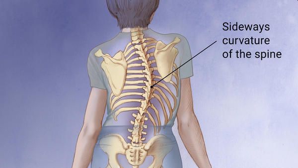
When we talk about scoliosis, we immediately understand that we are referring to a curvature of the spine, but it should be noted that not all column curvatures are, says the orthopaedic in Delhi.

Curves derived from postural "bad habits", reflex curvatures caused by pain or contractures, or compensatory "scoliosis", in which the curvature occurs to compensate for other asymmetries, are not considered scoliosis.

Causes and symptoms of scoliosis

Depending on the type of scoliosis, these will have one or the other causes. As a general rule, scoliosis due to malformations is congenital, but can also manifest itself from other diseases. Despite this, most scoliosis is idiopathic, appears in childhood or adolescence, and its causes are unknown, explains the orthopaedic in Delhi.

Scoliosis, although congenital, is not detected in the new-born, but appears progressively in childhood and stabilizes when bone growth ends, after puberty, even increasing in old age if degenerative phenomena appear.

It should be noted that neither the postures adopted by the child, nor the intense practice of exercise increases the risk and that the greater the degree of curvature there is a greater volume of scoliosis in girls than in boys.

Studies show that scoliosis does not produce more back pain or more intense pain than patients who do not have it, at least in slight curvatures, nor that 'straightening' the spine leads to significant improvements in the quality of life of those who suffer it, except the merely aesthetic.

In severe curvatures it can present chronic back pain or in extreme cases even affect breathing or some organs, says the orthopaedic doctor in Delhi.

Scoliosis Treatment

Considering that scoliosis does not produce pain or relevant limitations - by itself and in degrees of slight curvature - the chosen treatment and its impact should be considered based on its consequences, that is, not choosing an overly aggressive treatment for a problem (scoliosis) whose effect is mainly aesthetic.

Depending on the degree of scoliosis, the indicated treatments (in order) are:
  • Non-specific exercise: It simply seeks to improve the muscular and cardiovascular state, it has positive effects in terms of prevention and evolution of back pain, which is why it is highly recommended for children both for scoliosis itself and for their general health. and to establish in the child that healthy habit.
  • Specific exercise: Traditionally it was prescribed with the idea of ​​developing muscles with the ability to reduce scoliotic curvature, but there is no evidence of its effectiveness in this regard, although it does exist when it comes to reducing the prescription of the brace.
  • Corset: Its only objective is to stop the development of the curvature, but its continued use can generate muscular atrophy. Although the materials of current corsets are much more flexible to be more comfortable, they are less effective than rigid ones. Even so, the observable changes in a patient with a brace and another without a brace are not very relevant, so its use is being rethought - especially in children.
  • Surgery: Only cases of scoliosis associated with other diseases or malformations need to be operated, so it would not make sense to submit a patient with idiopathic scoliosis to a surgical process since the risks and scars are considerable to obtain a merely 'aesthetic benefit. '. The surgery is aggressive and usually has relevant complications in 10.2% of the operated, some examples are; wound infections, respiratory or implant complications. In addition, scoliosis surgery involves loss of mobility of the spine or limitation of rotational movements, depending on the area of ​​the spine affected, explains the orthopaedic surgeon in Delhi.

Prevention

Although due to the origin of this 'disease' it is difficult to prevent it in most cases, there are certain recommendations to prevent it or not make it worse. The practice of exercises to strengthen the area, the practice of swimming (an exercise whose benefits for the back in general are well known), the correct postural hygiene, not abusing heels and visiting regularly a physiotherapist to assess our state and help us not aggravate scoliosis are the most common forms of 'prevention'.

Gait studies also play an important role in the diagnosis and prevention of scoliosis, especially in children and adolescents in whom it is still possible to act because they are in the growth phase, says the orthopaedic in Delhi.

Saturday 11 July 2020

WHY DOES THE KNEE CLICK?

Knee-Clicking

Clicking in the knee joint is a fairly common cause of patients turning to orthopaedic surgeon in Delhi. In some patients, these clicks are accompanied by pain, but in most cases, these clicks do not cause any extraneous sensations. Some patients may have a history of trauma to the knee joint, in most cases, patients do not remember anything like this. One fine day, the patient simply notices that his knee has begun to click. Treatment is not necessary in all such cases, but in some of them it can be indicated.

The vast majority of patients who notice clicks in the knee do not experience pain, so do not worry about it. There are a number of interesting scientific studies that show that patients who have their knees clicked become very anxious. They begin to worry that clicks in the knee joint will lead to rapid wear of the articular cartilage and degenerative damage to the knee joint. In fact, clicks in the knee joint in the vast majority of cases are caused by the movement of normal anatomical structures in the area of ​​the knee joint. And this means that you will not have any osteoarthritis as a result. A number of studies have shown that clicks can be caused by gas in the knee joint. This is not dangerous. Also, clicks can be caused by movement of joint fluid in the joint cavity, explains the orthopaedic in Delhi.

Now let's talk about situations where clicks in the knee joint are accompanied by pain. Consider a number of the most common causes of this phenomenon.

WHAT CAUSES CLICKS IN THE KNEE JOINT?

Our knee joints are formed by three bones - the lower end of the femur, the upper end of the tibia and the patella. These bones are stabilized and held next to each other by a large number of ligaments. Movement in the knee joint is ensured by the coordinated work of powerful large muscles and tendons. Between the ends of the bones forming the knee joint are menisci, which are a kind of shock absorbers.

Any of the structures described may be damaged due to physical exertion, injury, or even due to normal wear and tear. Clicks in the knee joint can be caused by damage to almost any of the anatomical structures described above, explains the orthopaedic in Dwarka.

The most common causes of clicks and other extraneous mechanical sensations in the knee joint:
  • Chondromalacia: This is a condition characterized by degeneration of the articular cartilage, such as the patella. As a result of degeneration, the normal smooth surface of the articular cartilage becomes uneven. When the uneven articular surface of the patella moves along the corresponding articular surface of the femur, clicks, noise, or crunching occur.
  • Meniscus tears: between the joint ends of the femur and tibia in the knee joint are two menisci. The menisci resemble the letter “C” in shape and protect the articular surfaces of the bones that form the knee joint. When the meniscus is damaged, its torn fragment can cause clicks in the knee joint with certain movements.
  • Cartilage defects: the bones forming the knee joint are covered with a thick and smooth layer of “articular cartilage”. Injuries and certain diseases, such as dissecting osteochondritis, can lead to defects in the cartilage layer. Such a damaged articular surface becomes uneven, as a result of which various mechanical sensations may appear, including clicks in the knee joint. This is not the most common cause of painful clicks in the knee joint.
  • Dislocation of the patella. Dislocations of the patella are much more common than is commonly thought. If you have tucked up your foot and felt a click in the knee joint during any kind of sports, it is highly likely that the patella was dislocated. The patella is located in the groove on the anterior surface of the femur. Ideally, it should be located exactly in the center of this furrow. Sometimes the patella moves in the furrow in one direction or another, and sometimes it completely leaves it. Dislocation of the patella or a violation of its normal biomechanics is one of the most common causes of clicks in the knee joint. This is a very complex problem that requires a full examination by an orthopaedic doctor in Delhi who specializes in the problems of small tracking and instability of the patella.
  • Syndrome of a pathological synovial fold: the synovial fold is a fold of the inner lining of the knee joint. Many people have such folds, but they are relatively infrequent cause clicks and pain in the knee joint. Over time or as a result of repeated loads, these folds can thicken and become inflamed, causing painful clicks in the knee joint.
  • Osteoarthritis: This is a degeneration or "thinning" of the articular cartilage. Normal articular cartilage is an incredible smooth and even tissue. As osteoarthritis progresses, the surface of the cartilage becomes more and more uneven. Sometimes such cartilage can even peel off from the underlying bone, resulting in a cartilage defect. Any irregularities in the articular surfaces can cause clicks in the knee joint during movement.

If such simple means as a pressure bandage, muscle stretching and temporary limitation of physical activity do not bring the desired effect, it makes sense to consult an orthopaedic doctor in Dwarka.
There are other, less common causes of clicks in the knee joint. The causes described above usually cause not only clicks, but also pain in the knee joint. If clicks in your knee are accompanied by pain, we recommend that you consult your doctor.

For many of us, these clicks are completely painless. Sometimes their causes remain unknown. In many situations, these clicks can be considered a "norm" that does not require any treatment, says the orthopaedic in Delhi.

Tuesday 7 July 2020

Wrist and Elbow Fractures: Symptoms and Treatment

Elbow Wrist Fracture

To examine a distal radius fracture, your orthopaedic in Delhi will need to take an x-ray of the wrist. An open fracture is when the bone breaks through the skin. If the fracture extends into the joint, it is called an intra-articular fracture. Fractures that do not enter the joint are called extra-articular fractures.

Distal radius fracture treatment in Delhi includes the instantaneous measure of stabilizing the arm and placing an ice pack. It does this to keep the injury from getting even worse and to reduce swelling and control pain. A non-surgical option is casting if the bone is in good condition. The orthopaedic in Delhi may find it essential to straighten (or reduce) the bone to properly align it. Surgery is essential when the bone must be reduced through an incision. This is known as an open reduction, and these procedures generally require orthopaedic implants such as bone plates, pins, and / or bone screws to hold the bone parts in place.

Scaphoid fracture of the wrist

The scaphoid bone is one of several small bones in the wrist. It is located on the side of the thumb of the wrist where flexion occurs, just above the radius of the bone. When the scaphoid bone fractures, you will have pain, reduced wrist movement, swelling, bruising, and tenderness.

Most scaphoid fractures occur when falling with an outstretched hand. To identify a scaphoid fracture of the wrist, the physician will need to take radiographs to assess the displacement. However, several times, a break in this part does not appear immediately. If your orthopaedic doctor in Delhi suspects that the scaphoid is fractured, she will often apply a wrist splint for two weeks, and will come back sometime to repeat the analysis and possibly an MRI.

Scaphoid fracture treatment in Delhi depends on the precise location of the rupture. The fracture near the thumb usually heals for a few weeks with protection. If the fracture is more complex, the doctor may apply a cast to the wrist and monitor healing. Tears in the middle part of the scaphoid are more difficult to heal due to the limited blood supply.

The orthopaedic in Dwarka will generally suggest surgery for these types of scaphoid fractures. Your orthopaedic surgeon in Delhi will create a small incision and insert metal orthopaedic implants, to hold the bone in place when it heals. In rare cases, a bone graft is essential. Regardless of whether surgery is essential or not, you may need to wear the splint or cast for up to 6 months.

Elbow fractures

The bony aspect of the elbow that extends from the bone of the ulna arm is called the olecranon. This bulge is located under the skin with a slight protection against muscles or soft tissues. The elbow joint is made up of 3 bones, and these allow you to bend and straighten like a hinge. The humerus is the upper arm bone, the ulna is the lower arm bone, and the radius is the lower arm bone on the thumb side. When the elbow is injured, either by breaking one of these bones or by breaking a ligament that connects one bone to another, it can be painful, stiff and unstable.

Elbow fractures occur from a direct hit, such as being hit with a baseball bat, or indirectly from landing on an outstretched arm with the elbow locked in a straight line. Symptoms of an elbow fracture are bruising, sudden severe pain, swelling, inability to straighten the elbow, pain with movement of the joints, numbness in one or more fingers, and tenderness. To identify an elbow fracture, the orthopaedic doctor in Dwarka must look at the injury and take an x-ray of the joint.

Fracture treatment in Dwarka depends on the level of the injury. Some elbow fractures only require a sling, splint, or cast and conservative measures, while others require surgical intervention. If the bones are out of position, or if there are pieces of bone going through the skin, surgery will be required. The surgery is performed by expert orthopaedic surgeon in Dwarka using special orthopaedic instruments.

Typically, your orthopaedic surgeon in Delhi will make an incision over the back of the elbow and hold the bone parts and other structures together with wires, pins, braces, bone screws, and / or sutures. Once the surgery is done, a cast will be applied over a period of time and activity will be limited to allow for healing, says the orthopaedic in Delhi.

Wednesday 1 July 2020

COXARTHROSIS (OSTEOARTHRITIS OF THE HIP JOINT)

Coxarthrosis

Osteoarthritis, or arthritis due to wear and tear, is a common disease that occurs in people mainly middle-aged and elderly. According to rough estimates, in 2011 in the United States 29 million people suffered from this disease. Any joint of the body can be affected, however, the joints that bear the load are most often affected, including hip.

Osteoarthrosis of the hip joint or coxarthrosis in Latin - is characterized by the appearance of pain in the joint and limitation of movements. Patients may find it difficult to cope with everyday activities, such as tilting to tie shoelaces on boots, getting up from a chair, or taking short walks.

Osteoarthrosis progresses with time, so the sooner you start treatment, the less this disease will affect the way you live. There is no radical way to cure osteoarthritis, however, there are many ways to support therapy that will help you cope with pain and remain an active person, says the orthopaedic in Delhi.

HIP ANATOMY
Hip Anatomy
The hip joint is a spherical joint. Its "nest" is called the acetabulum, which is part of the pelvic bone. The second part of the joint is the spherical head of the femur.

The articular surfaces of the bones forming the hip joint are covered with a smooth tissue called articular cartilage. Cartilage provides unobstructed gliding of bones relative to each other.

Inside, the joint cavity is lined with a thin membrane called synovia. In a healthy hip joint, synovia produces a small amount of synovial fluid, which serves as a lubricant for articulating surfaces and improves their mobility.

WHAT IS COXARTHROSIS?

Osteoarthrosis is a degenerative joint disease that most often occurs at the age of 50 years and older, although it can also occur at a younger age, says the orthopaedic in Delhi.

With coxarthrosis, the articular cartilage of the hip joint gradually wears out. At the same time, its surface becomes uneven, it becomes thinner and the distance between the bone surfaces of the articulating bones decreases. As a result, the cartilage wears out completely and the bone surfaces begin to come into contact with each other. In order to somehow compensate for the loss of cartilage, on the periphery of the articular surfaces, the bone begins to grow and forms bone growths - osteophytes.

Coxarthrosis develops slowly and the associated pain syndrome progresses just as slowly.

CAUSES OF COXARTHROSIS

Coxarthrosis does not have any one specific reason, however, there are several factors in which the development of the disease is more likely:
  • Age
  • Family history of osteoarthritis
  • Previous hip injury
  • Obesity
  • Incorrect formation of the hip joint in the prenatal period - dysplasia of the hip joint

Even if you do not have the risk factors listed above, your likelihood of developing osteoarthritis still exists.

SYMPTOMS OF THE DISEASE

The most common symptom of coxarthrosis is pain. Pain usually develops and progresses slowly, but sometimes a sudden development of pain is possible. Joint pain and stiffness can be most pronounced in the morning, after prolonged sitting or a period of rest. Over time, the pain becomes more frequent, remains at rest and begins to bother the patient at night. Other symptoms include:
  • Pain in the groin and thigh radiating to the buttock or knee
  • Pain aggravated by intense physical exertion
  • Hip stiffness that makes normal walking and joint movement difficult
  • Sensation of joint blocking and crunching during movements associated with the presence of free fragments of cartilage and other tissues in the joint that interfere with normal movements.
  • Reduced range of motion, affecting the patient’s gait and causing lameness
  • Worse pain in rainy cold weather

CONSERVATIVE TREATMENT OF COXARTHROSIS

There is no radical way to cure coxarthrosis, but there are many ways to maintain therapy that can help you cope with pain and remain an active person.

As with degenerative damage to other joints, the initial treatment for coxarthrosis is conservative. The orthopaedic doctor in Delhi can recommend a variety of treatment options.

Lifestyle change. Some changes in your daily lifestyle will help protect your hip joint and slow down the progression of the disease.
  • Minimization of those types of activity that increase pain, for example, climbing stairs.
  • Replacing high-intensity loads (for example, running and tennis) with less intense ones (swimming and cycling).
  • Weight correction, which will reduce the load on the hip joint, thereby reducing the severity of pain and improving its function.
Physiotherapy. It includes special exercises that help increase the range of motion and flexibility of the joint, as well as strengthen the muscles surrounding the joint. The orthopaedic doctor in Delhi and physiotherapist will help you choose the individual exercise program that best suits your needs and lifestyle.

Supporting aids. Using canes, crutches, or walkers will help increase your mobility and make you more independent.

Drug therapy. If the pain interferes with your daily life and you cannot cope with it by other means, the doctor may recommend that you add medications to the treatment.
  • Over-the-counter analgesic can effectively help reduce the severity of pain in coxarthrosis. However, like any other drug, it can cause side effects and interact with other drugs that you take. We advise you to discuss possible side effects with your doctor.
  • Nonsteroidal anti-inflammatory drugs (NSAIDs) help reduce pain and inflammation, and most of them are also OTC drugs.
  • Corticosteroids are powerful anti-inflammatory drugs that can be administered orally or injected directly into the joint.

SURGICAL TREATMENT OF COXARTHROSIS

With severe pain and impaired joint function, when conservative treatment is ineffective, the orthopaedic doctor in Dwarka may recommend surgery.

Osteotomy. The operation consists in crossing the upper end of the femur or acetabulum and reorienting them in order to remove the load from the damaged part of the joint. In the treatment of osteoarthritis of the hip, this operation is rarely used.

Superficial arthroplasty. This operation consists in removing damaged cartilage and bone tissue of the acetabulum and replacing them with an artificial component. At the same time, the femoral head is not removed, and its articular surface is replaced by a smooth metal cap.

Total arthroplasty. This operation involves the complete replacement of the hip joint with artificial metal, plastic or ceramic components.

With total hip replacement in Delhi, the femoral head and acetabulum are replaced by artificial components.

Complications. Complications are possible in any surgical operation, and the orthopaedic surgeon in Delhi will do everything necessary to minimize their risk. The most common complications are:
  • Infection
  • Excess bleeding
  • Thrombosis
  • Hip dislocation
  • Difference in limb length
  • Blood vessel damage

RECOVERY AND REHABILITATION

After any operation on the hip joint, a rehabilitation period follows. The duration of this period depends on the nature of the operation performed.

Your orthopaedic in Delhi may recommend physiotherapy, which will help restore muscle strength and range of motion in the hip joint. After the operation, you will be using a cane, crutches, or walkers for some time.

In most cases, surgery can reduce joint pain and return you to an almost normal daily life.

The Revolution of Robotic Knee Replacement Surgery

Introduction Robotic knee replacement surgery has emerged as a groundbreaking advancement in the field of medical science. This innovative...