Showing posts with label orthopaedic surgeon in west Delhi. Show all posts

Thursday, 19 May 2022

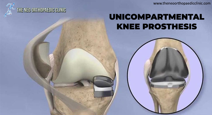
UNICOMPARTMENTAL KNEE PROSTHESIS


During knee replacement surgery in Delhi, the damaged bone and cartilage are covered with metal and plastic components. In a unicompartmental knee replacement (also called a “partial” knee replacement), only part of the knee is covered. This procedure is an alternative to total knee replacement for patients whose disease is limited to just one area of ​​the knee.

Because partial knee replacement is done through a smaller incision, patients generally spend less time in the hospital and return to normal activities sooner than patients undergoing total knee replacement.

ADVANTAGES OF PARTIAL KNEE REPLACEMENT

Several studies show that most patients who are suitable candidates for the procedure have good results with unicompartmental knee replacement in Delhi.
The advantages of partial knee replacement over total knee replacement include:

  • Faster recovery;
  • Less pain after surgery;
  • Less blood loss;

DISADVANTAGES OF PARTIAL KNEE REPLACEMENT

The disadvantages of partial knee replacement compared to total knee replacement include:

  • Slightly less predictable pain relief;
  • Potential need for more surgery. For example, a total knee replacement in Delhi may be necessary in the future if arthritis develops in the parts of the knee that were not replaced;

SURGERY CANDIDATES

If your osteoarthritis has advanced and non-surgical treatment options are no longer relieving your symptoms, orthopaedic in Dwarka may recommend knee replacement surgery in West Delhi. In order to be a candidate for unicompartmental knee replacement, your arthritis must be limited to one compartment of your knee. In addition, if you have any of the following characteristics, you may not be eligible for the procedure:

  • Inflammatory arthritis;
  • Significant knee stiffness;
  • ligament damage;

With proper patient selection, modern unicompartmental knee replacements have demonstrated excellent mid- and long-term results in younger and older patients.

YOUR SURGERY

A partial knee replacement operation typically lasts between 1 and 2 hours.

Partial knee replacement. There are three basic steps in the procedure:

  • Prepare the bone. Your orthopaedic surgeon in Dwarka will use special saws to remove cartilage from the damaged compartment of your knee;
  • Position the metal implants. The removed cartilage and bone are replaced with metallic coatings that recreate the joint’s surface. These metal pieces are typically held in the bone with cement;
  • Insert a spacer. A plastic insert is placed between the two metal components to create a smooth gliding surface;

COMPLICATIONS

As with any surgical procedure, there are risks involved with a partial knee replacement. Your orthopaedic surgeon in West Delhi will discuss each of the risks with you and take specific steps to help prevent potential complications.

Although rare, the most common risks include:

  • Blood clots. Blood clots in the leg veins are a common complication of knee replacement surgery. Blood clots can form in the deep veins of the legs or pelvis after surgery. Blood anticoagulants such as low molecular weight heparin and aspirin can help prevent this problem. Newer medications, such as rivaroxaban (Xarelto), may also be prescribed by your orthopaedic doctor in Dwarka, depending on your needs;
  • Infection. After surgery, an infection may occur in the skin over the wound or deep into the wound. An infection can happen while you are in the hospital or after you go home. You will be given antibiotics before the start of your surgery and these will be continued for about 24 hours afterwards to prevent infection;
  • Nerve or vessel damage. Although it rarely happens, nerves or blood vessels can be injured or stretched during the procedure;
  • continued pain;
  • Risks of anesthesia;
  • Need for additional surgery;

RECOVERY

Hospital discharge. Patients with partial knee replacement generally experience less postoperative pain, less swelling, and have easier rehabilitation than patients undergoing total knee replacement. In most cases, patients go home 1 to 3 days after the operation. Some patients go home on the day of surgery.

Weight support. You will begin putting weight on your knee immediately after surgery. You may need a walker, or crutches for the first few days or weeks until you feel comfortable enough to walk without assistance.

Rehabilitation exercise. A physiotherapist in Dwarka will give you exercises to help maintain your range of motion and restore your strength.

Tuesday, 17 May 2022

Patellar Instability or Patellar Dislocation


What is Patellar Instability or Patellar Dislocation?

The patella, formerly known as the kneecap, is the front bone of the knee, responsible for transmitting the strength of the thigh muscles. In some situations, this bone can move out of its normal location, called patellar instability or patellar dislocation, explains the orthopaedic in Delhi.

If the patellar dislocation occurred for the first time, it is called a primal patellar dislocation. From the second episode, it is called recurrent patellar dislocation.

What are the symptoms of patellar instability?

When a patellar dislocation occurs, there is severe pain and an inability to mobilize the knee. It is possible to see and feel that the patella has moved out of place.

Most of the time, the patella comes back into place on its own almost immediately. Rarely, a doctor needs to put it in place with a knee extension maneuver.

After a patellar dislocation, there may be a feeling of insecurity with the knee, even without the patella clearly moving out of place. This sensation is called a patellar seizure. It is a very uncomfortable symptom, which can interfere with normal activities, says the orthopaedic in Dwarka.

How and why does patella dislocation occur?

Patellar dislocation can occur from trauma, such as a blow or twist to the knee, or without trauma, in a common movement of the joint.

Some people have knee features that favor patellar dislocation. Among the main ones are:

  • High patella
    • The patellar tendon is longer, which makes the patella rest on the knee in a higher position, decreasing the bony socket at the beginning of knee flexion.
  • Increased patellar tilt
  • Femoral trochlea dysplasia
    • The groove on the femur where the patella fits can be shallower than normal, completely flat, or even convex.
  • Increase of the “Q” angle
    • “Q” angle is formed by the direction of traction of the thigh musculature and the direction of traction of the patellar tendon.
  • Patients with valgus knees (knees in, or in “X”)
  • Ligament laxity is also more predisposed.

What is the medial patellofemoral ligament?

The medial patellofemoral ligament is the structure that prevents dislocation of the patella. When the patella is displaced, it is injured or loosened.

When the patellar dislocation is treated without surgery, what is expected is that this ligament will heal. In the surgical treatment of patellar dislocation, reconstruction of the medial patellofemoral ligament is performed in most cases, explains the orthopaedic surgeon in Delhi.

Learn more about patellar dislocation treatments below.

How is patellar instability diagnosed?

The diagnosis of patellar instability is made through a careful assessment of the patient’s clinical history and physical examination, complemented with imaging tests.

The main test to be evaluated is magnetic resonance imaging, which shows indirect signs of dislocation, injury to the medial patellofemoral ligament, and the anatomical changes that favor instability. In addition, MRI is essential to look for cartilage lesions. Other tests, such as radiographs in special positions and computed tomography, are useful for evaluating the shape of the knee and predisposing factors, states the orthopaedic in west Delhi.

How is patellar instability treated without surgery? In what situation is he indicated?

In the case of a patient with an episode of patellar dislocation, both non-surgical and surgical treatment are possible. The decision for one or the other must be individualized, after a detailed discussion between the patient and the orthopaedic surgeon in Dwarka.

Non-surgical treatment involves immobilization for a period, followed by rehabilitation focused on exercises to strengthen and control the thigh and hip muscles. The goal of successful non-surgical treatment is the absence of new episodes of dislocation and patellar apprehension, the feeling of discomfort or buckling caused by instability, explains the orthopaedic surgeon in Dwarka.

In which cases is surgery indicated?

Situations that indicate treatment with patellar dislocation surgery are:

  • Recurrent episodes of dislocation (recurrent patellar dislocation)
  • Association with cartilage injuries
  • Symptoms of apprehension getting in the way of normal activities

Patients with a single episode of dislocation, although they can be treated without surgery, can also opt for surgical treatment. Surgery has the advantage of a lower chance of re-displacement or seizure symptoms for activities, says the orthopaedic surgeon in west Delhi.

What are patellar instability surgeries like?

According to the orthopaedic surgeon in Delhi, there are several procedures available for patellar instability, which are chosen according to the characteristics of each patient, and there may be a combination of procedures. This concept of individualized treatment is known as à la carte treatment, influenced by the French school.

These are the most common procedures performed for patellar dislocation.

  • Reconstruction of the medial patellofemoral ligament
    This ligament is the main restrictor of patellar dislocation, and its reconstruction is indicated in almost all cases. It is a graft from the patient’s own tendon to remake the ligament.
  • Lateral release (or release)
    Release of structures that hold the patella on the side or outside, when there is excess tension. Can be done openly or arthroscopically (video surgery)
  • Tibial tuberosity osteotomy
    A cut is made in the tibial bone to reposition the point where the patellar tendon attaches. This transfer allows for patellar realignment or patellar height correction.
  • Trochleoplasty
    Correction of the shape of the femoral trochlea, the groove where the patella rests on the femur.
  • Treatment of cartilage injuries
    When cartilage injuries also exist, these may also need specific treatment.

How is the postoperative period and rehabilitation?

Postoperative care depends on the technique used. They usually involve a period of support with crutches and a knee brace. However, from the beginning, it is already allowed to put the foot on the floor and remove the immobilizer to move the knee in most situations. Rehabilitation includes restoring knee mobility and restoring strength and control of the musculature of the thigh, hip, and trunk.

Wednesday, 20 April 2022

Everything About Knee Replacement

THE KNEE JOINT

Joints are the areas where bones meet, and movement occurs. The knee joint is made up of the femur above and the tibia below. The two bones are separated by cartilage that acts as a cushion and allows movement.

REASONS FOR SURGERY

The reasons for total knee replacement surgery in Delhi are: severe pain, loss of mobility, or deformity of the knee. Symptoms may be due to osteoarthritis, rheumatoid arthritis, or trauma among others.

Osteoarthritis, commonly called "wear and tear," is the most common cause for a total knee replacement.

ABOUT KNEE REPLACEMENT

The knee joint is made up of the ends of the thigh bone (femur) and the shin bone (tibia). These bones normally slide over each other with ease because they are covered by soft cartilage. If an injury damages the cartilage or is worn away by arthritis, for example, it can make the joint ache or stiff.

Generally, a new knee joint improves mobility and decreases pain, although your new knee will not be able to bend as much as a normal knee joint.

Depending on the condition of your knee joint, they will replace part or all of your knee joint. A total knee replacement is more common.

Artificial knee pieces can be made of metal and / or plastic, and a knee replacement can last up to 20 years.

WHAT ARE THE ALTERNATIVES?

Knee replacement surgery in Delhi is generally recommended only if nonsurgical treatments, such as physical therapy and exercise, taking medication, or using physical support devices such as a cane, no longer help decrease pain or improve mobility.

Alternative surgical procedures include arthroscopy in Delhi (if the arthritis is not very severe) or osteotomy (in which the leg bones are cut and put back). You may have already had these procedures before your knee replacement.

The surgeon will explain your options.

PREPARING FOR A KNEE REPLACEMENT

The orthopaedic surgeon in Delhi will explain how to prepare for the operation. For example, if you smoke, they will ask you to stop smoking, as this increases your risk of chest and wound infection, which can delay your recovery.

Typically, you must stay in the hospital for about five days, and the surgery is performed under general anesthesia. This means that you will be asleep during the operation. Otherwise, if you prefer, the surgery can be performed under epidural or spinal anesthesia. This type of anesthesia completely numbs from the waist down, and you will remain awake during the operation.

If you are going to have general anesthesia, you will be asked to fast. This means that you should not eat or drink, normally, for about six hours before general anesthesia. However, it is important to follow the instructions of your anesthetist.

In the hospital, the nurse can check your heart rate and blood pressure and do a urine test.
Your surgeon will explain to you what will happen before, during, and after the procedure, and any pain you may have. This is your opportunity to understand what will happen, and it may be helpful to prepare questions about the risks, benefits, and other alternatives to the procedure. This will help you stay informed so that you can give your consent if you are asked to sign a consent form to carry out the procedure.

You may be asked to wear compression stockings on your unaffected leg to prevent blood clots from forming in your veins (deep vein thrombosis, DVT). You may need an injection of a blood-thinning medicine called heparin in addition to, or instead of wearing, compression stockings.

WHAT HAPPENS DURING A KNEE REPLACEMENT?

Generally, a knee replacement in Delhi takes about two hours.

The orthopaedic in Delhi will make a single cut (10 to 30 cm long) in the front of your knee. You will push the kneecap to the side to reach the knee joint. The surgeon will remove the worn or damaged surfaces from the end of the femur and the top of the tibia. Typically, he will remove the anterior cruciate ligament and may remove the posterior cruciate ligament. For support, the best orthopaedic in Dwarka will not remove the collateral ligaments. It will shape the surfaces of the femur and tibia to fit the artificial knee joint and then fit the new joint over both bones.

Sometimes the back of the kneecap is replaced with a piece of plastic. This is known as patella lining.
After placing the new joint, the surgeon will close the wound with stitches or clips and cover it with a bandage. The surgeon will place a tight bandage on your knee to help minimize swelling.

WHAT SHOULD I EXPECT AFTER


You will need to rest until the anesthesia wears off. After epidural anesthesia, you may not be able to feel or move your legs for several hours.

You may need pain relievers to ease any discomfort when the anesthesia wears off.

You may have an intermittent compression pump attached to special pads on your lower legs for the first day or so. By inflating the cushions, the pump encourages healthy blood circulation and helps prevent a DVT. You can also have a compression stocking on your unaffected leg. This helps maintain circulation.

A physiotherapist in Dwarka (a movement and mobility specialist) will visit you daily to guide you through exercises that will help you recover.

You will stay in the hospital until you can walk safely with the help of a cane or crutch. When you can go home, you will need to ask someone to drive you.

Before you go home, the nurse will give you recommendations for caring for your knee and a date for your follow-up appointment.

How long it takes for the sutures to disappear will depend on the type used in the surgery. However, for this procedure they usually go away in about six weeks. Nonabsorbable sutures and clips are removed 10-14 days after surgery.

RECOVERING FROM KNEE REPLACEMENT SURGERY

If necessary, you can take an over-the-counter pain reliever, for example acetaminophen or ibuprofen. Follow the instructions in the patient information leaflet that comes with your medicine, and if you have questions, ask your pharmacist.

Physical therapy exercises are an indispensable part of your recovery, so it is essential that you continue to do them for at least two months.

You will be able to move around your house and go up and down stairs. For a few weeks, some everyday activities, such as shopping, will be difficult for you to do. You may need to use a cane or crutches for about six weeks.

You may be asked to wear compression stockings at home for several weeks.

When resting, raise your leg and support your knee to help prevent leg and ankle swelling.

Depending on the type of work you do, you may be able to go back to work after six to 12 weeks.

Follow your surgeon's recommendations for driving. You should not drive until you are sure that you can brake in an emergency without discomfort.

WHAT ARE THE RISKS?

Knee replacement surgery in West Delhi is a common and generally safe procedure. However, in order to make an informed decision and consent, you must be aware of the possible side effects and risk of complications associated with this procedure.

Side effects

These are the unwanted, though mostly temporary, effects of successful treatment; for example, feeling dizzy as a result of general anesthesia.

Your knee will hurt and be swollen for up to six months.

You will have a scar on the front of your knee. You may not have sensation in the skin around the scar. This may be permanent, but it should get better in two years.

Complications

Complications are problems that occur during or after the operation. Most of the people are not affected. Possible complications from any operation include unexpected reactions to anesthesia, excessive bleeding, or clot formation, usually in a vein in the leg (DVT).

Complications specific to knee replacement are rare, but include:

  • Wound or joint infection Antibiotics are given during and after surgery to prevent this complication.
  • Unstable joint. The knee joint may loosen and may require surgery to correct it.
  • Damage to blood vessels or nerves. It is usually mild and temporary.
  • Scar tissue. Scar tissue formation can limit movement. You will likely need another surgery to correct it.


Friday, 3 September 2021

HIP FRACTURES: ALL YOU NEED TO KNOW


The femur is the longest and strongest bone in the human body. For its fracture to occur, the effect of a sufficiently high force is necessary. One of these reasons could be, for example, a car accident.

The long, straight part of the femur is called the diaphysis. The fracture can occur at any part of it. Such fractures almost always require surgical treatment.

TYPES OF HIP FRACTURES

Depending on the energy of the injury, the nature of femoral fractures can vary greatly. Fragments may remain in their normal position (stable fractures) or significantly displaced (displaced fractures). The skin in the area of ​​the fracture may be intact (closed fracture) or it may be damaged, and the fracture may communicate with the external environment (open fracture).

Fractures are referred to by orthopaedic doctor in Delhi according to various classification systems. Hip fractures are classified according to:

  • Fracture localization (femur diaphysis is divided into thirds – distal, middle and proximal)
  • The nature of the fracture (the fracture line can be located in different ways: transversely, obliquely, etc.)
  • Damage to the skin and soft tissues in the area of ​​the fracture.

The most common types of hip shaft fractures are:

Transverse fracture. In this fracture, the line runs horizontally across the long axis of the thigh.

Oblique fracture. The fracture line is located at an angle to the axis of the thigh.

Spiral fracture. The fracture line is located in a spiral, as if surrounding the diaphysis of the thigh. The mechanism of such fractures is twisting along the long axis of the thigh.

Comminuted fracture. With such fractures, three or more bone fragments are formed. In most cases, the number of bone fragments is proportional to the severity of the traumatic effect that caused the fracture.

Open fracture. In such cases, the bone fragment can perforate the skin, or there is an open wound in the fracture area that communicates with the fracture zone. Open fractures are often characterized by greater damage to the surrounding muscles, tendons, and ligaments. These fractures have the highest risk of complications, especially infections, and usually take longer to heal.

CAUSES OF HIP FRACTURES

Fractures of the femur in young people are often the result of some kind of high-energy impact. The most common cause of hip shaft fractures is car accidents. Other common causes are pedestrian collisions with a moving vehicle and falls from a height.

Low-energy injuries, such as falls from their own height, can cause hip shaft fractures in older people with poor bone quality.

SYMPTOMS AND DIAGNOSIS OF FRACTURES

A hip shaft fracture usually immediately results in severe pain in the affected area. The victim loses the ability to lean on the injured leg, the hip may look deformed – it may be shorter and take an uncharacteristic position.

HISTORY AND PHYSICAL EXAMINATION

The orthopaedic in Delhi must know the circumstances of your injury. This information will help the orthopaedic surgeon in Delhi assess the energy of the injury and the presence of possible collateral damage.

It is important that the orthopaedic in Dwarka knows about any comorbidities you have – hypertension, diabetes, asthma, or allergies. The doctor will also ask you if you smoke or take any medications.

After discussing with you the nature of the injury and history, the orthopaedic in Janakpuri will perform a thorough physical examination. In doing so, the doctor will assess your general condition and then the condition of the injured limb. In this case, the orthopaedic in West Delhi will pay attention to details such as:

  • Visible limb deformity (unusual angle, rotation, or shortening of the limb)
  • Damage to the skin
  • Hemorrhage
  • Perforation with bone fragments of the skin

After a visual examination, the best orthopaedic in Delhi palpates the thigh, lower leg and foot, not the subject of possible pathological changes, tension of the skin and muscles in the fracture area. Also, the doctor will assess the nature of the pulse on the foot. If you are awake, your doctor will evaluate sensitivity and movement in your lower leg and foot.

RADIATION RESEARCH METHODS

Radiation testing allows the doctor to obtain more detailed information about your injury.

Radiography. It is the most commonly used method for diagnosing bone fractures. It allows not only to see the fracture, but also to characterize its type and localization.

Computed tomography. If the doctor needs more information about the nature of the fracture than is shown on the x-ray, the doctor may prescribe a CT scan. Sometimes the fracture line is very thin and almost invisible on radiographs. CT can help visualize these fractures more clearly.

TREATMENT OF HIP FRACTURES

CONSERVATIVE TREATMENT

Most hip shaft fracture treatment in Delhi require surgical intervention and rarely can be treated conservatively. So, the method of plaster immobilization is sometimes used to treat hip fractures in young children.

SURGERY

The timing of the operation. Most hip fractures are best operated within the first 24 to 48 hours after injury. Sometimes the operation is postponed due to the presence of life-threatening conditions or the need to stabilize the patient’s condition. To reduce the risk of infection in open fractures, patients are given antibiotics right after hospitalization. During the operation, open wounds, tissues and bone fragments are treated from contamination.

During the waiting period between admission to the hospital and surgery, the orthopaedic surgeon in Dwarka may temporarily fix your leg with a cast or skeletal traction. This allows you to maintain a more or less optimal position of the fragments and the length of the limb.

Skeletal traction is a system of blocks and weights with which bone fragments are held in one position. It allows not only to achieve the correct position of the fragments, but also to stop the pain syndrome.

External fixation. During such an operation, metal wires or rods are inserted into the femur above and below the fracture site, which are fixed to an external fixation device. This allows you to keep the fragments in the correct position.

External fixation is most often used as a method of temporary stabilization of a fracture in patients with multiple injuries, whose condition does not allow performing a more traumatic operation of internal fixation of the fracture. The second stage in such cases is performed after the patient’s condition has stabilized. In some cases, the external fixator is left on until the fracture is completely healed, but this is not common.

Intramedullary osteosynthesis. Today it is the most commonly used method of internal fixation of hip shaft fractures. In this case, special metal rods are used, which are inserted into the medullary canal of the femur. The rod passes through the fracture zone and holds the fragments in the correct position.

An intramedullary nail is inserted into the medullary canal from the side of the hip or knee joint. Above and below the fracture site, the rod is locked with screws to exclude mobility in the fracture area.

Intramedullary rods are usually made of titanium. They come in various lengths and diameters to fit most of the thigh bones.

Plates and screws. In such operations, the bone fragments are repositioned first, i.e. returning them to their normal position, after which the fragments are fixed from the side of the outer surface of the bone with a metal plate and screws.

This method is used when intramedullary osteosynthesis is not possible, for example, when the fracture line extends to the hip or knee joint.

RECOVERY AND REHABILITATION

Most fractures of the femoral shaft will heal within 3-6 months. Sometimes, for example, with open or comminuted fractures, as well as in smokers, it takes longer.

PAIN RELIEF

Pain after injury or surgery is a natural component of the healing process. Your doctor and nurses will do whatever is necessary to reduce pain and make your recovery more comfortable.

Various medications are usually used to relieve pain after an injury or surgery. These are paracetamol, non-steroidal anti-inflammatory drugs, muscle relaxants, opioids and topical drugs. In order to optimize the analgesic effect and reduce the patient’s need for narcotic analgesics, these drugs are often used in combination with each other. Some of these drugs can have side effects that affect your ability to drive or engage in other activities. The doctor will definitely tell you about the possible side effects of the drugs prescribed to you.

LOAD

Many doctors recommend starting movements in the joints of the operated limb as early as possible, but you need to load the leg when walking only in this way and only when and as your doctor permits.

In some cases, almost full loading is allowed immediately after the operation, but sometimes this is possible only after the first signs of fracture union appear. Therefore, we recommend that you strictly follow all the instructions of your orthopaedic surgeon in West Delhi.

You will need to use crutches or walkers when walking.

PHYSIOTHERAPY

After surgery, the muscles in the area of ​​the fracture are likely to be significantly weakened, so exercises to help restore muscle strength are very important during the rehabilitation process. Physiotherapy in Dwarka will restore normal muscle strength and joint mobility. It can also help you cope with postoperative pain.

A physiotherapist in Dwarka will likely start working with you while you are still in the hospital. He will also teach you how to use crutches or walkers correctly.
Saturday, 23 May 2020

HIP ENDOPROSTHETICS

Hip-Endoprosthetics

Pain in the hip joint is most often the result of osteoarthritis and can seriously affect your ability to lead a full and active lifestyle. Hip osteoarthritis is called coxarthrosis in medicine.

Endoprosthesis of the hip joint can help relieve pain and return to normal life. Over the past 20 years, thanks to the introduction of new materials and techniques into practice, the results of endoprosthetics operations have significantly improved.

Endoprosthesis of the hip joint is becoming more and more prevalent as the world's population is aging. At the moment, hip replacement surgery in Delhi is the most commonly performed in the world.

HIP ANATOMY
Hip-Anatomy

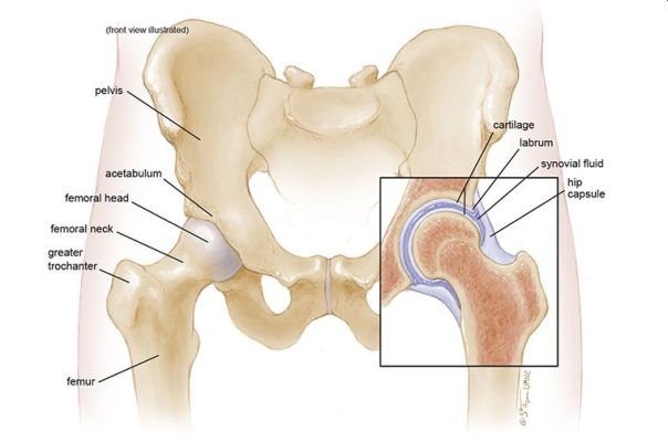
The hip joint is spherical in structure, so movements in it are possible in many planes. The joint is formed by the acetabulum, forming, as it were, a deep bowl and the head of the femur, which has the shape of a ball, explains the orthopaedic in Delhi.

The femoral head is connected to its main part (diaphysis) using a short portion of the bone called the femoral neck. Strong and thick muscles and tendons surround the joint. The surfaces of the acetabulum and femoral head are covered with articular cartilage. The thickness of the articular cartilage is about half a centimeter in large joints. The articular cartilage is a hard and smooth material covering the bones in the joint area. The articular cartilage allows the bones coated with it to glide smoothly relative to each other without being damaged. The color of the articular cartilage is white and shiny.

The joint is surrounded by a dense waterproof capsule, inside which a special fluid is produced that lubricates the mating surfaces. The bones in the joint hold tight ligaments and muscles together. The design of the hip joint provides extremely high mobility while maintaining satisfactory stability. The powerful muscles around the joint allow us to move for a long time in an upright position, and also, if necessary, accelerate when running and jumping. Important nerves and blood vessels also pass around the joint.

WHEN CAN ENDOPROSTHETICS REQUIRED?

The main indications for hip replacement in Delhi are arthrosis of the hip joint (coxarthrosis), fracture of the femoral neck, aseptic necrosis of the femoral head.

With arthrosis, degenerative changes in the articular cartilage occur, which ultimately leads to cartilage wear. Bone growths (osteophytes) form around the joint.

Due to the deterioration of the cartilage, a decrease in its thickness, a significant decrease in smoothness, as well as a change in the shape of the articular surfaces, the friction in the joint increases, which leads to pain and a progressive violation of the movements in the joint.

Aseptic necrosis of the femoral head is another cause of destruction of the hip joint. With this disease, the femoral head loses blood supply and actually collapses. The shape of the femoral head changes, the bone tissue making up the head is resorbed.

The articular surfaces of the acetabulum and the femoral head cease to correspond in shape, pain and impaired movement in the joint appear. The causes of the disease can be previous hip dislocations, birth injuries, prolonged treatment with corticosteroids, as well as some infections.

The main goal of replacing the joint in any of the degenerative diseases with an artificial one is to reduce pain and return movements. To do this, damaged surfaces are replaced with artificial ones, as a result of which the smoothness and painlessness of movements in the joint returns.

Fracture of the femoral neck is also an indication for joint replacement surgery in Delhi.

In case of fractures of the femoral neck, the blood supply to the head is disturbed, in connection with which its gradual destruction occurs.
Fracture fusion in these conditions is impossible, hip replacement surgery in Delhi is the only way to activate the patient and return him to everyday activity.

PREPARATION FOR HIP JOINT REPLACEMENT

The decision about the operation is made by the orthopaedic doctor in Delhi together with the patient. After clarifying the medical history, the doctor performs a thorough clinical examination to measure the current range of motion, the level of pain, the patient’s functionality. During the examination of the patient, the orthopaedic surgeon in Delhi examines the radiographs, as well as the data of CT and MRI studies.

A thorough and complete medical examination before surgery is also required. This is done in order to minimize the risk of complications during hip replacement surgery in Delhi. If a long-term operation or hemoglobin level of the patient is expected to be below normal values, a blood transfusion may be required after or during the operation. Mandatory prophylaxis of thromboembolic complications.

TYPES OF ENDOPROSTHESES

There are several main types of endoprostheses - cementless and cement.

Cemented endoprostheses are held in the bone using a special cement that fixes the metal to the bone. The surface of cementless prostheses is made in such a way that the bone tissue grows into it over time, due to which the prosthesis is held in the bone. In order for the endoprosthesis to grow, the bone is treated with special tools.

Both types of fixation of endoprostheses are widely used in medical practice. Also, in some cases, a combination may be used where, for example, the acetabular component (cup) is fixed with cement, and the femoral component (leg) is cementless. The decision about whether to use a cement or cementless endoprosthesis is made by the orthopaedic surgeon in Delhi based on the age, lifestyle of the patient and the quality of his bones.

The endoprosthesis consists of two main parts.

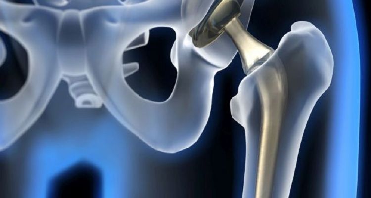
The acetabular component (cup) replaces the articular surface of the acetabulum. The shell of the acetabular component is made of metal, inside of which is placed a plastic or ceramic insert that is directly in contact with the femoral component.

The femoral component replaces the head and neck of the femur, usually made entirely of metal. In some designs of the endoprosthesis, the head may be made of ceramic.

Endoprosthetics can be total when both components are replaced, and unipolar. With unipolar endoprosthetics (hemiarthroplasty), only the femoral component changes. Hemiarthroplasty is usually performed for fractures of the femoral neck in elderly and debilitated patients.

With this type of endoprosthetics, the earliest verticalization of the patient is allowed, the very next day. This significantly reduces the risk of thromboembolic and hypostatic complications in elderly debilitated patients with femoral neck fractures. Equally important is the shorter operation time for hemiarthroplasty compared with total arthroplasty, which also reduces the risks during anesthesia and blood loss during surgery. Currently, our clinic uses modern cemented bipolar hip arthroplasty. A bipolar endoprosthesis is a modern type of unipolar prosthesis in which the head is double.

A similar design of the endoprosthesis increases the life of the prosthesis, increases its stability and range of motion.

MORE ABOUT HIP JOINT OPERATION

The orthopaedic surgeon in Delhi performs access to the hip joint, a skin incision is performed in the upper third of the thigh. After the hip joint is exposed, surgeons dislocate the worn femoral head from the acetabulum.

Then, a resection of the damaged head and neck of the femur is performed with a special electric saw.
Then, using special mills, the acetabulum is processed. During the treatment, the worn cartilage is completely removed and a hemisphere is formed into which the acetabular component will be implanted.

After the formation of the acetabulum, the surgeon fills the cavity with bone cement and establishes a suitable acetabular component. At this stage, the correct spatial orientation of the acetabular component at the right angle is important. This affects the life of the endoprosthesis and the likelihood of complications in the postoperative period.

After cement hardens and fixation of the acetabular component, the surgeon proceeds to the femur. At this stage, the bone canal of the femoral canal is developed with special rasps to the required size.

Next, cement is placed in the prepared canal in the femur and the femoral component is installed.

A head of the required size is selected and the femoral component is set into the acetabular.

After the orthopaedic surgeon in West Delhi checks the stability of the thigh and range of motion.

As soon as the surgeon is convinced that everything is set properly, the wound is sutured in layers. Drainages are established for a day. The patient is sent to a special ward in the postoperative ward, explains the orthopaedic in Delhi.

From the first day, rehabilitation of the patient begins.

Tuesday, 7 April 2020

Orthopaedic Implants - Pros and Cons

Orthopaedic Implants - Pros and Cons

It is now easy to treat serious bone problems with the start of technological developments and this was made possible with modern orthopaedic implants.

Although some time ago, diseases and bone fractures were not as common and were often seen in adults today, the situation has changed. These problems are now problematic for young players and dancers as well. But thanks to today's advancements, very sophisticated orthopaedic equipment, such as bone plates and orthopaedic bone screws, exists today with manufacturers of implants and orthopaedic instruments that can treat people with multiple bone fractures.

Orthopaedic surgeon in Delhi is performing fruitful surgeries and placing orthopaedic implants in their patients' bodies, thus relieving them of various bone disorders. In the case of severe fractures, these implants have proven to be very useful, as they have the ability to hold the fractured bones in place.

But if you are having this surgery for a bone plate and bone screw implantation, please read the following instructions to help you fully.

Precautions before opting for surgical implants: -
  • Consult your orthopaedic in Delhi before the operation and request tests before the surgery so that you do not develop sensitivity to the metal in your bones after the surgery. This test will decide if the screws and orthopaedic plates are appropriate for your skin and body or not. As some cases of severe skin reactions and wound infections have been reported by patients.
  • Next, be sure to opt for good-quality, non-toxic orthopaedic implants to protect yourself from any kind of inflammation after implantation.
  • Talk to your orthopaedic surgeon in West Delhi so that after surgery, you will have no difficulty making normal body movements, or you will have more problems than before.
  • If available, one can opt for biodegradable implants that possess properties and qualities almost similar to those of the original bone, to avoid the trauma of undergoing a second surgery to remove bone screws.
  • Since the bone plates and screws are made of metals such as titanium and its alloys, the risk of corrosion cannot be totally eliminated. So, the best way to handle these kinds of problems is to use the same screws and metal plates on one bone.
  • Orthopedic in Delhi suggest to don't strain after surgery and rest well because broken bones or ankles take long enough, sometimes many months, to heal and rehabilitate that joint.
  • You may experience severe pain due to bone screws and bone plates, while these ortho-surgical implants take you through the healing process. In that case, consult your orthopaedic in Dwarka directly and request pain medication instead of getting rid of these ortho implants.
The risks and precautions detailed above are not to dissuade anyone from enjoying the advantage of this miracle kit. It is just a tip that can help you really minimize the pain and suffering that comes from bone-related problems. Recovery from your infected part, joints and ankles can be quick when these orthopaedic implants are adjusted by taking essential safety measures and you get the most out of them.

Monday, 26 August 2019

ORTHOPAEDIC SURGEON AND TYPES OF ORTHOPAEDIC TREATMENTS

orthopaedics

Orthopaedics is the medical specialty that focuses on diseases and injuries of the musculoskeletal system in the human body. This complex system includes joints, bones, tendons, ligaments, muscles and nerves that allow one to move, work and be active.

An orthopaedic surgeon in Delhi treats patients of all ages, from new-borns with club foot to young athletes who require arthroscopy in Delhi and older people with arthritis. Most of us would have contacted him in case of an injury due to a bad fall or a car accident or an accidental torsion of one of our limbs, which would cause sprains or muscle tears.

Your orthopaedic surgeon

Orthopaedic surgeon in West Delhi treats musculoskeletal system problems. This involves:
  • Diagnosis of your disorder or injury.
  • Treatment with exercise, casting, medication, surgery or other options. Surgery performed by an orthopaedic surgeon in Dwarka with special tools and instruments.
  • Rehabilitation recommending physiotherapy in Dwarka or exercises to restore movement, function and strength.
  • Prevention with treatment plans and information to prevent injuries or delay the progression of a disease.
While orthopaedic in Delhi is familiar with all the characteristics of the musculoskeletal system, several orthopaedists specialize in certain areas, such as the foot and ankle, shoulder, spine, hip, hand, or knee. They can also choose to focus on specific fields such as trauma, paediatrics or sports medicine. Some orthopaedic in West Delhi may specialize in many areas.

Visit and medical treatment

Your orthopaedic surgeon in Delhi will take a history of your illness or injury and perform a physical exam. This can be followed by diagnostic studies, such as blood or X-ray tests or CT scans / MRI.

Depending on the diagnosis, the orthopaedic in Najafgarh develops the best treatment plan so that he can lead a functional and active life.

Non-surgical treatment

Orthopaedic in Janakpuri treats various musculoskeletal conditions without surgery using exercises, medications and other rehabilitation or alternative therapies.

For most orthopaedic injuries and diseases, there is more than one form of treatment. The orthopaedic in Tilak Nagar may recommend the option of surgical treatment in cases where non-surgical options do not help.

Surgical treatment

Orthopaedic surgeons perform various types of surgeries. The common procedures are:
  • Arthroscopy: A procedure that uses special cameras and orthopaedic equipment to visualize, diagnose and treat problems within a joint.
  • Fusion: A "welding" process by which bones are fused with internal devices (such as metal rods) and bone grafts to heal in a single solid bone.
  • Internal fixation: A method to keep broken bone pieces in a suitable place with metal plates, pins or bone screws while the bone is healing.
  • Joint replacement surgery in Delhi (partial, total and revision): When a damaged or arthritic joint is removed and replaced by an artificial joint called a prosthesis.
  • Osteotomy: The correction of bone deformity by cutting and repositioning bone.
  • Soft tissue repair: Repair of soft tissues, such as torn ligaments or tendons.

10 Common Orthopedic Problems and Their Warning Signs

Our bones, joints, muscles, and ligaments support almost everything we do, from walking and climbing stairs to exercising and simply getting...