Saturday 30 April 2022

10 Common Signs and Symptoms of Hip Wear


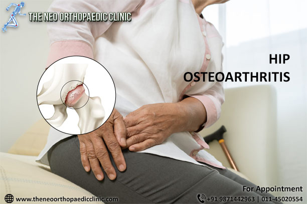
Hip wear (hip arthrosis) is a very frequent and limiting problem. It is the aging of articular cartilage that is responsible for free joint movement and pain-free. When the cartilage is worn out, the hip joint can no longer move freely and bear load, causing pain and movement restriction.

About 10% of the population over 45 years has hip wear with painful symptoms and almost 30% of the population has changes in hip wear on imaging tests such as radiography or MRI, says the orthopaedic in Delhi.

Common Symptoms

If you have a suspected hip wear, these are 10 most common Signs and Symptoms, which may indicate an evaluation with the hip surgery specialist. 

1. Pain in the groin area.

Pain in the groin region, or anterior region of the hip is perhaps the most common feature of the hip that is worn. This occurs by the characteristic of innervation of the joint that occurs by the same nerve roots that inners the groin region and anterior face of the thigh.

Pain in the groin area can occur because of other diseases, but it is very characteristic of the hip that has its cartilage worn out, states the orthopaedic in Delhi.

2. Stiffness of the hip joint. Loss of mobility.

One of the signs that a joint is worn is the loss of its function, which is precisely the ability to move the joint without pain.

When a joint is worn out, it ignites, and movement begins to cause pain. The result of this is that the patient himself begins to move the joint less as a way to protect himself from pain.

Thus, soft tissue structures such as capsule, muscles and tendons are retracted further reducing joint mobility, explains the orthopaedic doctor in Delhi.

3. Pain for lifting from low chairs or toilet.

Another striking feature of patients with hip arthrosis is the difficulty of getting up from low chairs and from the toilet.

This occurs because at the time of elevation, there is a sudden increase in load and pressure in the hip joint, which if worn out, will lead to a worsening of the pain, says the orthopaedic doctor in Delhi.

4. Claud gait, or “limp” gait.

The perfect functioning of the hip joint is essential for a balanced and pain-safe gait. When the hip is worn out, movement and change of loads on damaged cartilage can cause pain.

An immediate reflex is the decrease in the range of motion of the joint during gait and shortening of the pitch during the gait step on the worn hip.

All this gait movement in order to reduce the pain, ends up causing the “limp gait” or clauaudicante gait, explains the orthopaedic in Dwarka.

5. Pain to crouch and put on the shoes.

As much as it sounds like a simple activity, putting on simple shoes gets harder and harder for those who have hip arthrosis.

This occurs because the movement of putting on the shoes implies a large flexion of the hip and increased load on the joint, even if the patient is still.

A good alternative for patients who have hip arthrosis and pain to put on shoes, is to replace shoes with shoelaces with sneakers or shoes of the type “moccasin” that do not need to be tied, suggests the orthopaedic in Dwarka

6. Pain to go up and down stairs and to get in and out of the car.

This complaint is very common in patients with advanced hip arthrosis. With the progression of wear, pain is worse in activities with hip flexion with load and rotational movements, says the orthopaedic doctor in Dwarka.

Everyday activities such as going up and down stairs and getting in and out of the car get more difficult, requiring the support of the hands and the other member to be executed.

7. Feeling of locking, clicking, or crackling of the hip.

In many cases of hip wear, there may be detachments of cartilage fragments and inflammatory process in the joint (synovite).

These factors cause noises called clicking, or the famous “crek crek”, explains the orthopaedic doctor in Dwarka.

8. Decreased ability to walk and use supports

The hip is fundamental for a correct efficient and pain-free gait movement. Hip wear in a load area prevents perfect joint slippage and causes pain at the time of limb support.

This leads to a decrease in the patient’s ability to walk who needs to stop after a few steps for pain relief or the need to use supports such as crutches or walking, explains the orthopaedic in west Delhi.

9. Decrease in sexual activity.

Hip wear can reach many patients with active sex life. This can be a big problem because pain and limitation of movements can decrease the willingness to have sex (libido) or impair the sexual act due to the accentuated symptoms, says the orthopaedic in west Delhi.

10. Discouragement to carry out daily activities.

Hip wear is a progressive and limiting problem. It is very difficult to assimilate the loss of function and quality of life that it causes.

The constant pain and limitation for small daily activities greatly affects the psychological of patients. It’s very difficult to live with that.

When the pain is strong and the discouragement is very strong, it is good to remember that there is always the solution of the surgery. Look for a reliable specialist orthopaedic doctor in west Delhi

How many of these symptoms do I need to have to have surgery?

There is no specific number of symptoms that define the exact time of performing hip prosthesis surgery.

The higher the number of symptoms and the higher the intensity of them, the greater the chance of a hip replacement surgery in Delhi by a synthetic prosthesis.

Wednesday 20 April 2022

Everything About Knee Replacement

THE KNEE JOINT

Joints are the areas where bones meet, and movement occurs. The knee joint is made up of the femur above and the tibia below. The two bones are separated by cartilage that acts as a cushion and allows movement.

REASONS FOR SURGERY

The reasons for total knee replacement surgery in Delhi are: severe pain, loss of mobility, or deformity of the knee. Symptoms may be due to osteoarthritis, rheumatoid arthritis, or trauma among others.

Osteoarthritis, commonly called "wear and tear," is the most common cause for a total knee replacement.

ABOUT KNEE REPLACEMENT

The knee joint is made up of the ends of the thigh bone (femur) and the shin bone (tibia). These bones normally slide over each other with ease because they are covered by soft cartilage. If an injury damages the cartilage or is worn away by arthritis, for example, it can make the joint ache or stiff.

Generally, a new knee joint improves mobility and decreases pain, although your new knee will not be able to bend as much as a normal knee joint.

Depending on the condition of your knee joint, they will replace part or all of your knee joint. A total knee replacement is more common.

Artificial knee pieces can be made of metal and / or plastic, and a knee replacement can last up to 20 years.

WHAT ARE THE ALTERNATIVES?

Knee replacement surgery in Delhi is generally recommended only if nonsurgical treatments, such as physical therapy and exercise, taking medication, or using physical support devices such as a cane, no longer help decrease pain or improve mobility.

Alternative surgical procedures include arthroscopy in Delhi (if the arthritis is not very severe) or osteotomy (in which the leg bones are cut and put back). You may have already had these procedures before your knee replacement.

The surgeon will explain your options.

PREPARING FOR A KNEE REPLACEMENT

The orthopaedic surgeon in Delhi will explain how to prepare for the operation. For example, if you smoke, they will ask you to stop smoking, as this increases your risk of chest and wound infection, which can delay your recovery.

Typically, you must stay in the hospital for about five days, and the surgery is performed under general anesthesia. This means that you will be asleep during the operation. Otherwise, if you prefer, the surgery can be performed under epidural or spinal anesthesia. This type of anesthesia completely numbs from the waist down, and you will remain awake during the operation.

If you are going to have general anesthesia, you will be asked to fast. This means that you should not eat or drink, normally, for about six hours before general anesthesia. However, it is important to follow the instructions of your anesthetist.

In the hospital, the nurse can check your heart rate and blood pressure and do a urine test.
Your surgeon will explain to you what will happen before, during, and after the procedure, and any pain you may have. This is your opportunity to understand what will happen, and it may be helpful to prepare questions about the risks, benefits, and other alternatives to the procedure. This will help you stay informed so that you can give your consent if you are asked to sign a consent form to carry out the procedure.

You may be asked to wear compression stockings on your unaffected leg to prevent blood clots from forming in your veins (deep vein thrombosis, DVT). You may need an injection of a blood-thinning medicine called heparin in addition to, or instead of wearing, compression stockings.

WHAT HAPPENS DURING A KNEE REPLACEMENT?

Generally, a knee replacement in Delhi takes about two hours.

The orthopaedic in Delhi will make a single cut (10 to 30 cm long) in the front of your knee. You will push the kneecap to the side to reach the knee joint. The surgeon will remove the worn or damaged surfaces from the end of the femur and the top of the tibia. Typically, he will remove the anterior cruciate ligament and may remove the posterior cruciate ligament. For support, the best orthopaedic in Dwarka will not remove the collateral ligaments. It will shape the surfaces of the femur and tibia to fit the artificial knee joint and then fit the new joint over both bones.

Sometimes the back of the kneecap is replaced with a piece of plastic. This is known as patella lining.
After placing the new joint, the surgeon will close the wound with stitches or clips and cover it with a bandage. The surgeon will place a tight bandage on your knee to help minimize swelling.

WHAT SHOULD I EXPECT AFTER


You will need to rest until the anesthesia wears off. After epidural anesthesia, you may not be able to feel or move your legs for several hours.

You may need pain relievers to ease any discomfort when the anesthesia wears off.

You may have an intermittent compression pump attached to special pads on your lower legs for the first day or so. By inflating the cushions, the pump encourages healthy blood circulation and helps prevent a DVT. You can also have a compression stocking on your unaffected leg. This helps maintain circulation.

A physiotherapist in Dwarka (a movement and mobility specialist) will visit you daily to guide you through exercises that will help you recover.

You will stay in the hospital until you can walk safely with the help of a cane or crutch. When you can go home, you will need to ask someone to drive you.

Before you go home, the nurse will give you recommendations for caring for your knee and a date for your follow-up appointment.

How long it takes for the sutures to disappear will depend on the type used in the surgery. However, for this procedure they usually go away in about six weeks. Nonabsorbable sutures and clips are removed 10-14 days after surgery.

RECOVERING FROM KNEE REPLACEMENT SURGERY

If necessary, you can take an over-the-counter pain reliever, for example acetaminophen or ibuprofen. Follow the instructions in the patient information leaflet that comes with your medicine, and if you have questions, ask your pharmacist.

Physical therapy exercises are an indispensable part of your recovery, so it is essential that you continue to do them for at least two months.

You will be able to move around your house and go up and down stairs. For a few weeks, some everyday activities, such as shopping, will be difficult for you to do. You may need to use a cane or crutches for about six weeks.

You may be asked to wear compression stockings at home for several weeks.

When resting, raise your leg and support your knee to help prevent leg and ankle swelling.

Depending on the type of work you do, you may be able to go back to work after six to 12 weeks.

Follow your surgeon's recommendations for driving. You should not drive until you are sure that you can brake in an emergency without discomfort.

WHAT ARE THE RISKS?

Knee replacement surgery in West Delhi is a common and generally safe procedure. However, in order to make an informed decision and consent, you must be aware of the possible side effects and risk of complications associated with this procedure.

Side effects

These are the unwanted, though mostly temporary, effects of successful treatment; for example, feeling dizzy as a result of general anesthesia.

Your knee will hurt and be swollen for up to six months.

You will have a scar on the front of your knee. You may not have sensation in the skin around the scar. This may be permanent, but it should get better in two years.

Complications

Complications are problems that occur during or after the operation. Most of the people are not affected. Possible complications from any operation include unexpected reactions to anesthesia, excessive bleeding, or clot formation, usually in a vein in the leg (DVT).

Complications specific to knee replacement are rare, but include:

  • Wound or joint infection Antibiotics are given during and after surgery to prevent this complication.
  • Unstable joint. The knee joint may loosen and may require surgery to correct it.
  • Damage to blood vessels or nerves. It is usually mild and temporary.
  • Scar tissue. Scar tissue formation can limit movement. You will likely need another surgery to correct it.


Sunday 17 April 2022

Hip arthroscopy in athletes: eliminate exercise pain

Every athlete who suffers from hip pain usually faces a very similar journey: when the first symptoms appear, they start conservative treatment with anti-inflammatory drugs and painkillers. When the pain improves a little, they try to return to activity. After a little while, they are in pain again.

But when drug treatment no longer has any effect and the pain starts to impair sports practice or even the performance of simple routine activities, what can amateur and high-performance athletes do?

The first step is to seek an orthopedic in Delhi specializing in the hip, who will be able to perform a thorough evaluation. Only after that, treatment will start based on the cause of the injury, that is, try to treat the problem at its root instead of just treating the pain.

The imbalance of strong structures around the hip joint, such as tendons, ligaments and bursae are the main cause of hip pain in those who practice sport. This happens because the hip makes an important connection between the legs and the trunk. Any slight alteration can generate instability and injury.

While many athletes have no indication for surgery, for others, performing a surgical procedure, such as hip arthroscopy in Delhi, is the best option to restore quality of life, sports performance and prevent an injury from becoming chronic, or even progress to cartilage wear.

Hip arthroscopy for athletes

Problems such as hip injuries are closely linked to the practice of sports such as running, cycling, volleyball, tennis, football, dance, artistic gymnastics and other sports.

In the recent past, a large number of young people with hip pain during physical activity did not have a correct diagnosis and some cases progressed to wear and tear. The orthopaedic doctor in Delhi acted as a spectator of the worsening of the joint injury, without being able to interfere with the inevitable.

Today, it is known that many of these young athletes had a disease caused by a change in the fit of the frame, called femoroacetabular impingement.

Athletes with this type of hip injury are the main beneficiaries of hip arthroscopy surgery in Delhi.

Considering the limitations in sports performance imposed by the hip injury, the procedure is a great treatment option for femoroacetabular impingement with labrum injury, as it is capable of correcting the anatomy of the joint socket.

Today, there are several studies that demonstrate that the rate of return to sport after arthroscopy is above 90%. The rehabilitation phase after hip arthroscopy in athletes lasts about 6 months, and may vary for each case.


How hip arthroscopy works in athletes

Hip arthroscopy is a minimally invasive surgery, in which 1 to 2 cm incisions are made in the hip to place cameras and specific instruments used to repair cartilage and adjust the hip bones.

The performance of arthroscopy in athletes is indicated mainly for those who have excess bone in the hip joint, have pain and limitation of movement. These patients are usually adults between 20 and 40 years of age who practice physical activity.

Hip arthroscopy in athletes, when recommended, is a great solution to eliminate pain and return to physical exercise.

A common mistake made by many athletes is accepting hip pain as part of their routine and continuing to practice sports even without a proper diagnosis.

Many patients live with hip problems for years and seek treatment only when the pathology is preventing them from performing movements.

Having medical follow-up from a hip specialist orthopedic in Dwarka will ensure that the athlete has adequate treatment from the start, preventing the injury from progressing and requiring more invasive treatments.

Faced with the persistence of symptoms, look for a specialist orthopaedic surgeon in Delhi!

Friday 15 April 2022

Knee Replacement Surgery For Accute Knee Pain

Knee replacement surgery, also known as "knee arthroplasty," can help relieve pain and restore function to severely diseased knee joints. This procedure involves cutting out damaged bone and cartilage from the femur, tibia, and patella, and replacing it with an artificial joint (prosthesis) made of metal alloys, premium plastics, and polymers.

To determine if a knee replacement in Delhi is right for you, an orthopedic surgeon in Delhi evaluates your knee's range of motion, stability, and strength. The use of X-rays helps determine the degree of damage.

Your doctor can choose from a variety of knee replacement prostheses and surgical techniques based on your age, weight, activity level, knee size and shape, and overall health.

One of the most common reasons for knee replacement surgery in Delhi is severe pain caused by damage to the joint due to wear and tear from arthritis (osteoarthritis). Osteoarthritis can wear down the slippery cartilage that helps the knee joint move smoothly. An artificial knee joint has metal alloy caps for the femur and tibia, and high-density plastic to replace damaged cartilage.

Knee Replacement Surgery

In traditional knee replacement surgery in West Delhi, the surgeon makes a long incision in the center of the knee joint, through muscles, tendons, and ligaments to reach the knee joint. In total knee replacement surgery in Delhi, the surgeon will remove damaged tissue and rebuild the surface of the knee joint with artificial materials. By replacing damaged and worn knee surfaces, total knee replacement surgery can reduce pain, correct leg deformities, and help many patients return to normal activities. Knee replacement surgery, called total knee arthroplasty, involves replacing damaged structures in the knee joint with metal and plastic pieces to restore normal knee function and relieve chronic pain.

In partial knee replacement, damaged cartilage and bone are removed and replaced in only one affected part of the knee joint. Total knee arthroplasty requires the implantation of an artificial joint to replace damaged or diseased cartilage and bone in the femur, tibia or patella. The procedure involves removing damaged bone and cartilage from the femur, tibia, and patella and replacing them with artificial joints (prostheses) made of metal alloys, high-quality plastics, and polymers. During this procedure, plastic and metal inserts are used to replace bone and cartilage in all parts of the knee joint, including the medial, lateral, and patella.


Surgeons cover the ends of the bones that make up the knee joint with metal or plastic parts, or implant a joint-shaped prosthesis. In total knee arthroplasty, orthopaedic surgeon in Dwarka uses metal to resurface the end of the femur (femur) and the thigh bone, where the two bones meet to form the knee joint. During surgery, the surgeon will remove diseased bone and cartilage where the femur (femur) and lower leg (tibia) meet at the knee joint. Surgeons left the original ligaments, tendons, skin, muscles and most of the bone and simply covered the damaged cartilage in the knee with metal plating.

Artificial knee joints have metal alloy caps for the femur and lower leg and high-density plastic to replace damaged cartilage. Knee replacement surgery in Delhi can help patients whose knee or knee joint has been damaged by injury due to osteoarthritis, rheumatoid arthritis, or post-traumatic arthritis. Knee replacement surgery may be appropriate when medications and other treatments fail to control knee pain. People who think they need medication for pain relief should consider seeing a joint replacement surgeon (an orthopaedic surgeon in West Delhi with experience in knee replacements) to determine if surgery is the best option.

Therefore, it is important that the best orthopaedic in Dwarka who performs a minimally invasive quadriceps-sparing total knee replacement is not only an excellent orthopaedic surgeon, but also an expert knee surgeon in West Delhi and minimally invasive knee arthroplasty. Even experienced knee replacement surgeons perform far more procedures using traditional methods than using less invasive methods; we know that the more procedures you do, the more reliable the results will be. Often, partial replacements are only beneficial for young people with symptoms in one part of the knee.

Saturday 9 April 2022

Patellar Tendinitis


What is patellar tendinitis?

One of the most common knee tendinitis or knee tendinopathies are those that result from inflammation of the patellar tendon.

The patellar tendon attaches proximally to the lower pole of the kneecap (or patella). This sesamoid bone transmits the pulling force of the quadriceps muscle, allowing mobilization of the knee joint. Distally, the tendon insertion zone is located in the region of the anterior tuberosity of the tibia.

Both insertions can be the site of inflammation and pain in the anterior (or rarely lateral) region of the knee, explains the orthopaedic in Delhi.

Causes of patellar tendinitis

Knee tendinitis is an injury that is very often associated with high-intensity sports training, especially one that requires repeated pushing movements (“jumper’s knee”).

Therefore, knee tendinitis of this type occurs mainly in sports that involve jumping (volleyball, handball, basketball, etc.).

However, knee tendinitis can also develop in occasional exercisers or in patients who do not exercise at all, says the orthopaedic in west Delhi.

Symptoms of Knee Tendonitis

The main signs and symptoms are pain in the anterior region of the knee, which worsens when jumping or running and which is sometimes accompanied by edema (swelling).

Knee pain is sometimes so intense that it can cause lameness and difficulty walking, states the orthopaedic in Dwarka.

Diagnosis of patellar tendinitis

The diagnosis of patellar tendinitis is made clinically, namely the type and location of pain, the clinical history of practice of certain modalities, as well as the analysis of auxiliary diagnostic tests such as: Knee X-Ray, Knee Ultrasound and the Magnetic Resonance Imaging (MRI) of the knee.

In case of doubt about the diagnosis or difficulties in overcoming the symptoms, you should consult your orthopaedic doctor in Delhi.

Complications in patellar tendinitis

In more severe cases of patellar tendinitis, namely those with a longer duration, the chronic inflammation can lead to progressive weakening of the tendon, with the appearance of micro-tears and eventual evolution to a complete and total tear of the patella.

Is patellar tendinitis curable?

Yes, there is a cure, the prognosis depending on a series of factors such as the time of evolution and intensity of symptoms, athlete’s biotype / weight, knee morphology, type and intensity of the sport practiced, explains the orthopaedic in Dwarka.

Age also decisively affects recovery time, being obviously shorter in younger athletes.

Find out below how to treat patellar tendinitis.

Treatment of knee tendonitis (patellar)

The treatment of all knee tendinitis in general always involves conservative measures, such as resting, applying local ice, systemic or topical anti-inflammatory drugs (or remedies).

Physiotherapy is always an essential part of this treatment, allowing to speed up recovery and rapid return to sport.

Infiltration with PRP ‘s is considered in the literature as having a local analgesic effect and improving the regeneration process.

In cases that are very resistant to these treatments and that cause great functional disability, infiltration with corticosteroids should be carried out with consideration given the increased risks of tendon rupture, especially when applied multiple times.

SURGERY FOR TENDONITIS OF THE KNEE (PATELLAR)

Surgery (or operation) for the treatment of these types of pathologies is always a last resort, says the best knee surgeon in west Delhi.

The surgical procedure consists of debridement of the degenerated tissues, opening the lower pole of the patella and, if necessary, reinserting any areas of tendon rupture, explains the best knee surgeon in west Delhi.

Due to the risks of possible complications, namely the complete rupture of the tendon, postoperative recovery must always be very cautious, respecting the healing timings of the tissues involved and under the supervision of specialist orthopaedic surgeon in Delhi and Physiatry.

Monday 4 April 2022

Knee Arthroscopy For Knee Injuries

What is knee arthroscopy?

Knee arthroscopy is a minimally invasive procedure that allows access and treatment of injuries that affect the various structures of said joint. For this, 2 or 3 small incisions of less than one centimeter are made that allow access to the joint.

Most knee surgeries that do not involve the placement of a prosthesis are performed with a complete or partial approach with knee arthroscopy in Delhi. It is the technique of choice to address many injuries because it allows a better and greater visualization of the joint. In a non-aggressive way, all the points of the knee can be accessed to clean the cartilage, small perforations of the bone that lacks cartilage (microfractures), stabilize the cartilage and apply substances or elements that allow the cartilage to regenerate (plasma rich in platelets or stem cells).

Likewise, knee arthroscopy is also used as support for other open techniques, since it improves the diagnosis and prognosis of the patient's injury, as it is less aggressive.

The arthroscopy in Delhi can be performed under local, regional or general anesthesia, depending on the injury and the patient. The anaesthesiologist will decide the best method for the patient, as long as he suffers as little as possible.

Why is it done?

Knee arthroscopy is used to solve knee injuries. Thus, meniscus injuries are one of the most common pathologies and, thanks to arthroscopy, it is possible to preserve most of the menisci, since the resection is not complete but partial. Meniscal sutures and the possibility of transplanting the meniscus with knee arthroscopy are common techniques that allow better protection of the cartilage of the joint.

Another of the most dangerous injuries related to sports practice is the rupture of the anterior cruciate ligament. If this is injured, it causes instability in the knee that makes it impossible for the patient to perform practically any sport. If the instability continues, it can damage surrounding structures, such as the menisci and cartilage. Hence, it is necessary to reconstruct the anterior cruciate ligament with grafts, accessing the joint through arthroscopy.

On the other hand, cartilage injuries (chondroplasties, osteoarthritis or osteochondritis) are also very common. Preserving the cartilage will also preserve the joint, avoiding wear and tear on the knee.


What does it consist of?

The orthopaedic in Delhi will make the small incisions in the knee to be able to access it. He will first fill the knee joint with a sterile solution and remove any cloudy fluid. This way he will be able to see the joint clearly and in detail.

The orthopaedic in Dwarka will then insert the arthroscope (a very thin device with a camera on the end) into the knee. This device sends the images to the television monitor, so that the surgeon can see all the structures in detail. Through the other holes, the surgeon will introduce the surgical material that will allow him to address the injury and repair the damaged structures.

It is a procedure that usually does not last more than an hour. After that, the patient will be transferred to a rehabilitation room and will be able to leave the hospital after two hours, more or less.

Preparation for knee arthroscopy

Before surgery, the patient must undergo a complete physical examination so that the specialist can assess their health and any anomaly that may interfere with the arthroscopy. Likewise, the patient must inform the orthopaedic surgeon in Delhi of the medication he takes, so that he can tell him which ones he should stop taking before the intervention. Some complementary preoperative tests will also be carried out, such as magnetic resonance imaging, electrocardiogram or blood tests.

Care after the intervention

Recovery after arthroscopy is faster than conventional open surgery. However, the advice of the specialist must be followed so that the knee recovers correctly.

It is normal for the patient to experience swelling in the days after the intervention, so it is recommended that the leg be elevated during those first days after the surgery. Also, the application of ice will relieve pain and reduce inflammation.

The incisions should also be healed, keeping them clean and dry. The orthopaedic in West Delhi will tell the patient when they can shower or change the bandage.

On the other hand, shortly after the intervention, the patient must begin rehabilitation exercises with a Physiotherapist in Dwarka, who will establish a program appropriate to the patient and the injury. This will help restore motion and strengthen your knee muscles.

Alternatives to this treatment

The alternative to knee arthroscopy in West Delhi will be conventional open surgery, which is currently only used in more serious cases, in which a prosthesis must be placed. Any other technique will suppose a greater invasion in the knee and a worse postoperative period.


Sunday 3 April 2022

Understand how to treat osteoarthritis and live without pain


Pain located in the hip, which appears mainly during the day or after getting up from a long period of sitting, may be caused by osteoarthritis of the hip, explains the orthopaedic in Delhi.

It is a problem that generates pain and decreases the patient’s movements, directly affecting the routine and the performance of simple activities, such as crossing the legs, getting into the car, tying shoes, standing, walking and performing physical activities, says the orthopaedic in Delhi.

Many people live with this type of pain for a long time and wonder if hip osteoarthritis is curable. In this article you will understand how to treat and control this problem.

Is hip osteoarthritis curable? Understand how it happens

Osteoarthritis in the hip is a problem caused by the loss of cartilage in the joint, which ends up causing the pain generated by the friction of bones where cartilage should be.

This is because the cartilage present between the bones of all joints in the body does not have the ability to hurt. Thus, we can move all our joints, without noticing anything wrong.

The problem begins when a cartilage injury begins that causes the bones to come into contact with each other in the joint. As said, cartilage doesn’t hurt, but bones do. From this initial injury, a path of pain and wear begins, explains the orthopaedic in west Delhi.

In practice, the hip is formed by the junction of two bone structures:

  • Femur Head – It is the round part of the hip joint, formed by the femur bone, which is the thigh bone.
  • Acetabulum – It is the part of the pelvis cavity formed by the bones of the pelvis.

These joints fit perfectly and tightly, allowing for correct and completely pain-free movement of the hip. The problem is when this joint is affected by diseases that cause cartilage loss – as is the case with osteoarthritis., states the orthopaedic in west Delhi.

Know the symptoms of osteoarthritis in the hip

The symptoms of osteoarthritis of the hip tend to evolve over time and tend to start with mild, localized pain in the hip, says the orthopaedic doctor in Delhi.

As wear and tear on cartilage progresses slowly, initial symptoms are usually mild and worsen over time.

The pain tends to worsen with the effort used in simple activities such as standing and physical activities, improving only when the patient is at rest.

Faced with worsening and worsening of symptoms, the joint starts to block movement, preventing the patient from performing activities such as putting on shoes, crossing the legs or cutting nails.

In more advanced stages, there may be pain even at rest, says the orthopaedic doctor in Delhi.

After all, is osteoarthritis in the hip curable?

Osteoarthritis in the hip is a problem that, in and of itself, has no cure.

However, no desperation. There are treatments that can and should be performed in order to reduce pain and improve symptoms caused by cartilage damage.

The first step for anyone who wants to improve from hip pain is to correctly diagnose the cause of the pain. This will help determine the best treatment approach and, of course, the best outcome. The evaluation and trust in qualified professionals can make a patient stagnant in treatment, live again, says the orthopaedic in Dwarka.

Learn about the main ways to treat hip arthrosis:

1. Medicines

Patients who have severe, sharp pain may benefit from using anti-inflammatory drugs or corticosteroids in order to reduce inflammation quickly.

2. Changing habits

It is recommended that those who have joint problems develop some habits such as:

  • Avoid consumption of sugar, alcohol and saturated fats, as these types of food can increase inflammation and increase pain.
  • Reduce the physical activity that causes hip pain.
  • Keeping the body moving with physical activity that does not generate pain and has a controlled impact.
  • Reducing body weight helps not only to lessen the weight on the injured joint, but also to control the process of widespread inflammation caused by fat in the body.

3. Physiotherapy

Physiotherapy is done as part of treatment to reduce pain and reorganize body structures damaged by osteoarthritis.

It should be done with the guidance of a specialist physiotherapist, through analgesic and anti-inflammatory therapies, exercises that aim to improve joint lubrication, work on hip amplitude and function, as well as balance and body compensation techniques.

4. Exercises

It is recommended to practice physical activities that help to strengthen the thigh muscles and work on stretching the region.

Activities such as pilates, water aerobics and cycling should be part of the routine of people who suffer from osteoarthritis in the hip, in order to preserve movements and improve the frame.

But beware, each person must be evaluated individually. An activity that can do a lot of good for one type of person can accelerate hip wear and tear in another.

5. Hip Injection

Hip Injection is a technique that can be applied to the structures around or directly inside the joint, and it can have the function of reducing inflammation or improving the joint situation.

According to the objective, substances such as corticosteroids (to reduce inflammation) and hyaluronic acid (to improve joint lubrication and environment) are injected.

The type of injection must be done according to the medical advice of the specialist orthopaedic doctor in West Delhi, according to previous analysis and treatment objective for each patient.

6. Hip Replacement Surgery

Surgery for osteoarthritis in the hip should only be performed under the guidance of a specialist orthopaedic surgeon in Delhi. It is recommended when other treatments do not help to control pain or when there is a well-defined intrinsic benefit.

The hip replacement surgery in Delhi consists of removing the two parts of the diseased bone and placing components of hip prostheses. In these cases, after recovering from anesthesia, while still under observation, the patient begins to walk with a walker, continuing the treatment with a physical therapist.

Osteoarthritis in the hip is treatable

Osteoarthritis in the hip has no cure, but it has treatment that should be used to reduce and control pain, regain joint mobility and return the patient’s quality of life, says the orthopaedic in Delhi.

With medical guidance, it is possible to control symptoms and identify the best way to lead a pain-free life without limitations due to osteoarthritis in the hip.

Living with pain is not normal. Look for a specialist orthopaedic doctor in Delhi to treat hip arthrosis and resume your routine activities.

The Revolution of Robotic Knee Replacement Surgery

Introduction Robotic knee replacement surgery has emerged as a groundbreaking advancement in the field of medical science. This innovative...