The long bones are part of the skeleton of the limbs. Through them, large-amplitude movements are performed, such as current gestures of daily life (the bones of the thoracic limb) as well as those necessary for walking (the bones of the pelvic limb). The diaphysis of the long bones are frequently exposed to trauma leading to fractures, says the orthopaedic in Delhi.
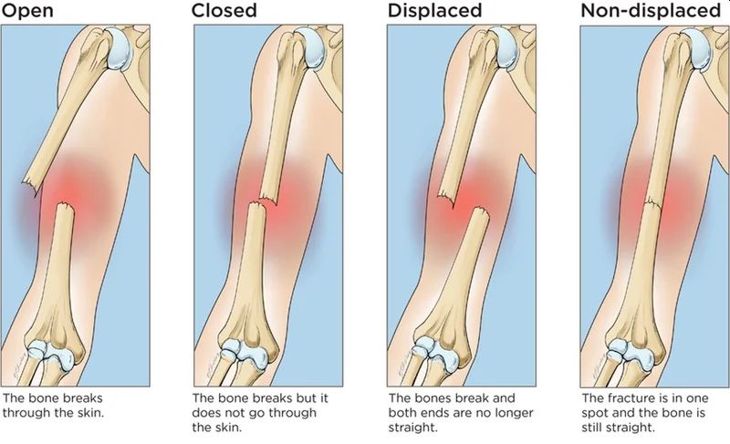
Fractures represent a discontinuity or interruption in a bone as a result of trauma.
Humeral shaft fractures are common between the neck and the humeral supracondylar region. Often the location of the fracture is on average 1/3.
Humeral shaft fractures are common in adults and much less common in children and the elderly. They can also be found during a difficult labour at birth, with the attempt to release the arm, also called an obstetric fracture.
In the case of humeral diaphyseal fractures, the mechanism of production is more frequently indirect compared to the direct mechanism which is much less common. The indirect mechanism determines the fracture with spiroid trajectory (torsion mechanism) or with short and transversal oblique trajectory, in the latter case there is bending by falling on the hand or elbow, explains the orthopaedic in Dwarka.
Symptoms
Following a fracture, general and local signs are found.
The general signs consist of agitation, anxiety, pallor and sometimes the state of shock can be found, especially in important accidents. Impairment of the general condition occurs more frequently in lower limb fractures, in open fractures in both the upper and lower limb, in polytraumas when other visceral injuries occur, explains the orthopaedic surgeon in Delhi.
Local signs of fracture may be signs of probability and certainty.
The probability signs are:
- fixed point pain accompanied by functional impotence. At the moment of the fracture, the patient has a very strong local pain, which subsequently diminishes, persisting a background pain that worsens when the fractured segment is mobilized. Due to the pain, the patient tends to keep the affected limb immobilized, installing functional impotence
- swelling of the area accompanied by deformity of the region, then bruising at the fracture
- local deformity
- vicious position by moving fragments and shortening the segment.
Signs of certainty confirm the presence of the fracture. These are:
- abnormal mobility
- the presence of bone crackling
- disruption of bone continuity
- in-transmissibility of movements - the impression of a movement of the distal fracture segment is not transmitted to the segment located proximal to the fracture due to the interruption of the bone lever.
In the conditions of an incomplete fracture (cracks) there are no signs of certainty of the fracture but only signs of probability says the orthopaedic surgeon in Dwarka.
Diagnosis and complications
Clinical examination and radiography of the humerus establish the diagnosis of certainty. Also, the radiograph at this level can specify if it is a pathological bone fracture. Often humerus is the seat of metastasis to bone spurs visceral neoplasms (breast cancer, lung cancer, kidney cancer, etc.)
A complication is immediate radial nerve paralysis, which is impaired in Santa twisting the humerus. The so-called "swan neck hand" appears. Another less common immediate complication is damage to the vessels that irrigate the humerus.
The most common late complication is osteoarthritis that develops progressively.
A vicious callus is formed which is well tolerated and does not cause functional disorders, only when there is an angle over 20-30 degrees accompanied by a significant gap and a shortening of more than 3 cm.
Treatment
The treatment is mainly conservative at the orthopaedic clinic in Delhi. It consists in reducing the movements under the action of the weight of the arm and of the external manoeuvres and the immobilization in a plastered thoraco-brachial apparatus with the arm next to the body.
If the reduction cannot be achieved, a plastered brachio-antebrachial hanging device can be used, which will favour the reduction in time (continuous traction effect). This device is maintained for 2 maximum 3 weeks and then is replaced with a thoraco-brachial device for another 21 days, says the orthopaedic doctor in Delhi.
In the case of fractures that cannot be reduced, with interposition, in transverse fractures, those on pathological bone, surgical treatment is indicated.
In oblique fractures, osteosynthesis is recommended by the orthopaedic in Delhi with brooches or rods. In transverse fractures, it is preferable to perform osteosynthesis with the compaction plate.
In the case of radial nerve palsy, the fracture is corrected and then the nervous phenomena recede. If no signs of regeneration appear in 6-8 weeks, neurolysis is indicated.
Pseudoarthrosis is treated by plaque osteosynthesis and according to the type of pseudoarthrosis by the contribution of spongy autograft.


No comments:
Post a Comment